Friday, April 16, 2010

Gout also known as gouty arthritis is a metabolic disease marked by monosodium urate deposits that cause red, swollen, and acutely painful joints. Gout can affect any joint but mostly affects those in the feet, especially the great toe, ankle, and midfoot. Gout is a medical condition that usually presents with recurrent attacks of acute inflammatory arthritis (red, tender, hot, swollen joint). It is caused by elevated levels of uric acid in the blood. The uric acid crystallizes and deposits in joints, tendons, and surrounding tissues. Gout affects 1% of Western populations at some point in their lives. Gout is caused by an increased level of uric acid in the blood, salts of which are deposited in the joints. It mostly occurs in middle-aged men and almost always involves pain at the base of the great toe. Gout may result from a primary metabolic disturbance or may be a secondary effect of another disease, as of the kidneys.Gout is treated with drugs to suppress formation of uric acid or to increase elimination of uric acid. Patients who receive treatment for gout have a good prognosis. 
The final, unremitting stage of the disease (also known as tophaceous gout) is marked by persistent painful polyarthritis. An increased concentration of uric acid leads to urate deposits in cartilage, synovial membranes, tendons, and soft tissue, called Tophi/tophus . Tophi/tophus form in the fingers, hands, knees, feet, ulnar sides of the forearms, pinna of the ear, Achilles tendon and, rarely, in such internal organs as the kidneys and myocardium. Renal involvement may adversely affect renal function. 

Causes for Gout/Gouty Arthritis 
Hyperuricemia is the underlying problem of gout, Although the underlying cause of primary gout is unknown, it appears to be linked to a genetic defect in purine metabolism that causes overproduction of uric acid (Hyperuricemia), retention of uric acid, or both. 
Secondary gout develops during the course of another disease, such as obesity, diabetes mellitus, hypertension, polycythemia, leukemia, myeloma, sickle cell anemia, and renal disease. Secondary gout can also follow treatment with such drugs as hydrochlorothiazide or pyrazinamide. 

Complications for Gout/Gouty Arthritis 
Potential complications include: Renal disorders such as renal calculi Circulatory problems, such as atherosclerotic disease, cardiovascular lesions, stroke, coronary thrombosis, and hypertension Infection that develops when occur tophi ruptures and nerve entrapment. 

Treatment for Gout/Gouty Arthritis 
Gout/Gouty Arthritis management has three goals: Stop the acute attack. Treat hyperuricemia to reduce urine uric acid levels. Prevent recurrent gout and renal calculi. 
Treatment for an acute attack: 
Bed rest; immobilization and protection of the inflamed, painful joints; and local application of cold. Analgesics, such as acetaminophen, relieve the pain associated with mild attacks. Acute inflammation requires nonsteroidal anti-inflammatory drugs or intramuscular corticotropin. 
Treatment for chronic gout involves 
Decreasing the serum uric acid level. Adjunctive therapy emphasizes avoidance of alcohol and sparing use of purine-rich foods. Weight reduction program decreases uric acid levels and eases stress on painful joints. In some cases, surgery may be necessary excised and drained tophi to improve joint function or correct deformities. 


Nursing Assessment 
Patient history Reveal that the patient has a sedentary lifestyle and a history of hypertension or renal calculi. report waking with pain in toe or another location in the foot.He may complain that initially moderate pain has grown and He may report accompanying chills and a mild fever. 
Inspection a swollen, dusky red or purple joint with limited movement. Maybe found tophi, especially in the outer ears, hands, and feet, In chronic stage of gout, the skin over the tophi may ulcerate and release a chalky white exudate or pus. 
Palpation may reveal warmth over the joint and extreme tenderness. The vital signs assessment may disclose fever and hypertension. If the patient has a fever, possible occult infection must be investigated. 

Diagnostic tests for Gout/Gouty Arthritis 
Needle aspiration of synovial fluid (arthrocentesis) or tophaceous material Serum uric acid X-rays 

Nursing Diagnosis
Common nursing diagnosis found in Nursing care plans for Gout/Gouty Arthritis Acute pain Activity intolerance Anxiety Deficient knowledge (diagnosis and treatment) Disturbed sleep pattern Impaired physical mobility Ineffective coping Risk for injury

Tuesday, April 13, 2010

Nursing Care Plans for Prostate Cancer. Prostate cancer is the most common neoplasm in males older than age 50; it’s a leading cause of male cancer death. Adenocarcinoma is the most common form; only seldom does prostate cancer occur as a sarcoma. Most prostate cancers originate in the posterior prostate gland, with the rest growing near the urethra. Malignant prostatic tumors seldom result from the benign hyperplastic enlargement that commonly develops around the prostatic urethra in older males. 
Slow-growing prostate cancer seldom produces signs and symptoms until it’s well advanced. Typically, when primary prostatic lesions spread beyond the prostate gland, they invade the prostatic capsule and then spread along the ejaculatory ducts in the space between the seminal vesicles or perivesicular fascia. When prostate cancer is fatal, death usually results from widespread bone metastases. 

Stage of Prostate Cancer 

  1. Stage A or I: Prostate cancer that is only found by elevated PSA and biopsy, or at surgery for obstruction. It is not palpable on DRE. It is usually found accidentally during surgery for other reasons, such as BPH, usually curable, especially if it is a relatively low Gleason grade. 
  2. Stage B or II: can be felt on rectal examination and is click to enlargelimited to the prostate. Other tests, such as bone scans or CT/MRI scans, may be needed to determine this stage, especially if the PSA Blood tests is significantly elevated or the Gleason grade is 7 or greater 
  3. Stage C or III: Cancer has already spread beyond the capsule of the prostate into localclick to enlarge organs or tissues, but has not yet metastasized or jumped to other sites 
  4. Stage D or IV: Cancer has already spread, first site usually pelvic and perivesicular lymph nodes and bones of the pelvis, sacrum, and lumbar spine 

Stage of Prostate Cancer

Causes for Prostate Cancer 
Risk factors for prostate cancer include age (the cancer seldom develops in males younger than age 40) and infection. Endocrine factors may also have a role, leading researchers to suspect that androgens speed tumor growth. 
Complications for Prostate Cancer 
Progressive disease can lead to spinal cord compression, deep vein thrombosis, pulmonary emboli, and myelophthisis. 

Nursing Assessment 
The patient’s history may reveal urinary problems, such as dysuria, frequency, retention, back or hip pain, and hematuria. The patient with these complaints may have advanced disease, with back or hip pain signaling bone metastasis. The patient usually has no signs or symptoms in early disease. Inspection may reveal edema of the scrotum or leg in advanced disease. During digital rectal examination (DRE), prostatic palpation may detect a nonraised, firm, nodular mass with a sharp edge (in early disease) or a hard lump (in advanced disease). 

Diagnostic tests for Prostate Cancer 
The American Cancer Society advises a DRE and a blood test to detect prostate-specific antigen (PSA) yearly for males age 50 and older with a life expectancy of at least 10 years. These screenings may be done for males at high risk of the disease beginning at age 40 to 45, depending on their risk factors. Blood tests may show elevated levels of PSA. Although most males with metastasized prostate cancer have an elevated PSA level, the finding also occurs with other prostatic disease, so the PSA level should be assessed in light of DRE findings. Transrectal prostatic ultrasonography may be used for patients with abnormal DRE and PSA test findings. Bone scan and excretory urography are used to determine the disease’s extent. Magnetic resonance imaging and computed tomography scanning can help define the tumor’s extent. 

Treatment for Prostate Cancer 
Therapy varies by cancer stage and may include radiation, prostatectomy, orchiectomy (removal of the testes) to reduce androgen production, and hormonal therapy with synthetic estrogen (diethylstilbestrol). Radical prostatectomy is usually effective for localized lesions without metastasis. A transurethral resection of the prostate may be performed to relieve an obstruction. Radiation therapy may cure locally invasive lesions in early disease and may relieve bone pain from metastatic skeletal involvement. It may also be used prophylactically for patients with tumors in regional lymph nodes. Alternatively, internal beam radiation may be recommended because it permits increased radiation to reach the prostate but minimizes the surrounding tissues’ exposure to radiation. If hormonal therapy, surgery, and radiation therapy aren’t feasible or successful, chemotherapy may be tried. Chemotherapy for prostate cancer (combinations of cyclophosphamide, doxorubicin, fluorouracil, cisplatin, etoposide, and vindesine) offers limited benefits. Researchers continue to seek the most effective chemotherapeutic regimen. 

Nursing Diagnosis 
Common nursing diagnosis found in Nursing Care Plans Prostate Cancer: 

  • Acute pain 
  • Anxiety 
  • Fear 
  • Impaired urinary elimination 
  • Ineffective coping 
  • Risk for infection Sexual dysfunction 


Nursing Key Outcomes 
The patient will voice increased comfort. The patient will report that he feels less anxious. The patient will verbalize concerns and fears related to his diagnosis. The patient will maintain an adequate urine output. The patient will demonstrate positive coping mechanisms. The patient will remain free from signs and symptoms of infection. The patient will acknowledge a problem in sexual function. 

Nursing interventions  
Provide encourage the patient to express his fears and concerns, including those about changes in his sexual identity, owing to surgery. Offer reassurance when possible. Give analgesics as necessary Administer ordered. Provide comfort measures to reduce pain. Encourage the patient to identify care measures that promote his comfort and relaxation. 

After prostatectomy 
Regularly check the dressing, incision, and drainage systems for excessive blood. Also watch for signs of bleeding (pallor, restlessness, decreasing blood pressure, and increasing pulse rate). Be alert for signs of infection (fever, chills, inflamed incisional area). Maintain adequate fluid intake (at least 2,000 ml daily). Give antispasmodics, as ordered, to control postoperative bladder spasms. Also provide analgesics as needed. Because urinary incontinence commonly follows prostatectomy, keep the patient’s skin clean and dry. 

After suprapubic prostatectomy 
Keep the skin around the suprapubic drain dry and free from drainage and urine leakage. Encourage the patient to begin perineal exercises between 24 and 48 hours after surgery. Allow the patient’s family to assist in his care and encourage them to provide psychological support. Give meticulous catheter care. After prostatectomy, a patient usually has a three-way catheter with a continuous irrigation system. Check the tubing for kinks, mucus plugs, and clots, especially if the patient complains of pain. Warn the patient not to pull on the tubes or the catheter. 

After transurethral resection 
Watch for signs of urethral stricture (dysuria, decreased force and caliber of urine stream, and straining to urinate). Also observe for abdominal distention (a result of urethral stricture or catheter blockage by a blood clot). Irrigate the catheter, as ordered. 

After perineal prostatectomy 
Avoid taking the patient’s temperature rectally or inserting enema or other rectal tubes. Provide pads to absorb draining urine. Assist the patient with frequent sitz baths to relieve pain and inflammation. 

After perineal or retropubic prostatectomy 
Give reassurance that urine leakage after catheter removal is normal and subsides in time. 

After radiation therapy 
Watch for the common adverse effects of radiation to the prostate. These include proctitis, diarrhea, bladder spasms, and urinary frequency. Internal radiation of the prostate almost always results in cystitis in the first 2 to 3 weeks of therapy. Encourage the patient to drink at least 2,000 ml of fluid daily. Administer analgesics and antispasmodics to increase comfort. 

After hormonal therapy 
When a patient receives hormonal therapy with diethylstilbestrol, watch for adverse effects (gynecomastia, fluid retention, nausea, and vomiting). Be alert for thrombophlebitis (pain, tenderness, swelling, warmth, and redness in calf). 

Patient teaching and home health guide 
Before surgery, discuss the expected results. Explain that radical surgery always produces impotence. Up to 7% of patients experience urinary incontinence. To help minimize incontinence, teach the patient how to do perineal exercises while he sits or stands. To develop his perineal muscles, tell him to squeeze his buttocks together and hold this position for a few seconds; then relax. He should repeat this exercise as frequently as ordered by the physician. Prepare the patient for postoperative procedures, such as dressing changes and intubation. If appropriate, discuss the adverse effects of radiation therapy. All patients who receive pelvic radiation therapy will develop such symptoms as diarrhea, urinary frequency, nocturia, bladder spasms, rectal irritation, and tenesmus. Encourage the patient to maintain a lifestyle that’s as nearly normal as possible during recovery. When appropriate, refer the patient to the social service department, local home health care agencies, hospices, and other support organizations.

Monday, April 12, 2010

Kidney Anatomy
Nursing Care Plans for Kidney Cancer, kidney cancer is rare, kidney cancer also called nephrocarcinoma; renal carcinoma, hypernephroma, and Grawitz’s tumor originate in the kidneys. Others are metastases from various primary-site carcinomas. Kidney cancers are classified by cell type. The three most commonly seen in the adult are renal cell carcinoma, transitional cell carcinoma, and sarcoma. 
Most kidney tumors are large, firm, nodular, encapsulated, unilateral, and solitary. They may affect either kidney; occasionally they’re bilateral or multifocal. Kidney cancer is twice as common in males as in females; it typically strikes after age 40. Kidney cancer can be separated histologically into clear cell, granular cell, and spindle cell types. Sometimes the prognosis is considered better for the clear cell type than for the other types; in general, however, the prognosis depends more on the cancer’s stage than on its type. 

Causes for Kidney Cancer
Although the cause of kidney cancer is unknown, some studies implicate several factors seem to predispose a person to kidney cancer. Smokers increase their risk to develop kidney cancer by 40%. A link also exists between kidney cancer and occupational exposure to cadmium (found in batteries), asbestos, some herbicides, benzene, and organic solvents, particularly trichloroethylene. Patients who receive regular hemodialysis may also be at increased risk. kidney cancer stage 


Complications for Kidney Cancer Related to metastasize to other sites

  • Respiratory problems from metastasis to the lungs, 
  • neurologic problems from brain metastasis 
  • GI problems from liver metastasis. 

Nursing Assessment Nursing Care Plans for Kidney Cancer
The patient may complain of hematuria and often a dull, aching flank pain. He may also report weight loss, although this is uncommon. Rarely, his temperature may be elevated. Palpation may reveal a smooth, firm, nontender abdominal mass. 

Diagnostic tests for Kidney Cancer

  • Renal ultrasonography 
  • Computed tomography scan 
  • Renal angiography 
  • Urography 
  • Nephrotomography
  • Kidney-ureter-bladder radiography. Additional relevant tests include liver function studies 


Treatment for Kidney Cancer

  • Radical nephrectomy. 
  • Radiation treatment is used only when the cancer has spread into the perinephric region or the lymph nodes or when the primary tumor or metastatic sites can’t be completely excised 
  • Chemotherapy 
  • Biotherapy with lymphokine (causes many adverse reactions) 
  • Hormone therapy, such as medroxyprogesterone and testosterone 


Nursing diagnosis 
Primary nursing diagnosis Nursing Care Plans for Kidney Cancer is altered urinary elimination related to renal tissue destruction, common nursing diagnosis found on Nursing Care Plans for Kidney Cancer: 

  • Acute pain 
  • Anxiety 
  • Fear 
  • Impaired physical mobility 
  • Ineffective breathing pattern 
  • Ineffective tissue perfusion: Renal 
  • Readiness for enhanced management of therapeutic regimen 
  • Risk for imbalanced fluid volume 


Nursing outcomes for nursing care plans for Kidney Cancer, Patient will:

  • Maintain urine specific agents within normal range 
  • Report increased comfort. 
  • Identify strategies to reduce anxiety. 
  • Express fears and concerns relating to his condition and prognosis. 
  • Maintain joint mobility and range of motion. 
  • Maintain ventilation. 
  • Communicate understanding of medical regimen, medications, diet, and activity restrictions. 
  • Maintain fluid balance. 


Nursing interventions for Kidney Cancer
Before surgery, assure the patient that the body will adequately adapt to the loss of a kidney. Administer prescribed analgesics as necessary. Provide comfort measures, such as positioning and distractions, to help the patient cope with discomfort. After surgery, encourage diaphragmatic breathing and coughing. Assist the patient with leg exercises, and turn him every 2 hours to reduce the risk of phlebitis. Check dressings often for excessive bleeding. Watch for signs of internal bleeding, such as restlessness, sweating, and increased pulse rate. Position the patient on the operative side to allow the pressure of adjacent organs to fill the dead space at the operative site, improving dependent drainage. If possible, assist the patient with walking within 24 hours of surgery. Provide adequate fluid intake, and monitor intake and output. Monitor laboratory test results for anemia, polycythemia, and abnormal blood chemistry values that may point to bone or hepatic involvement or may result from radiation therapy or chemotherapy Provide symptomatic treatment for adverse effects of chemotherapeutic drugs. Encourage the patient to express his anxieties and fears, and remain with him during periods of severe stress and anxiety. 

Patient Teaching And Home Healthcare Guide for Kidney Cancer
Tell the patient what to expect from surgery and other treatments. Before surgery, teach diaphragmatic breathing and effective coughing techniques, such as how to splint the incision Be sure the patient understands what medications are to be taken at home, their effects, and dosages. Explain follow-up information, such as when the physician would like to see the patient. Provide and arrange for a home visit from nurses if appropriate. Refer the patient and family to hospital and community services such as support groups Reinforce any postoperative restrictions. Explain when normal activity can be resumed. Make sure the patient understands the need to have ongoing monitoring of the disease. Annual chest x-rays and routine IVPs are recommended to check for other tumors. Emphasize and give understanding of the lifestyle choices that can aid in recovery e.g. Quit smoking, limit alcohol, eat more fruits, vegetables, and whole grains and less animal fat; exercise once you are able. Explain the possible adverse effects of radiation and drug therapy. Advise the patient how to prevent and minimize these problems. When preparing the patient for discharge, stress the importance of compliance with prescribed outpatient treatment.

Sunday, April 11, 2010

The choice of therapy depends on the type of hernia. For a reducible hernia, temporary relief may result from moving the protruding organ back into place. Afterward, a truss may be applied to keep the abdominal contents from protruding through the hernial sac. Although a truss doesn’t cure a hernia, the device is especially helpful for an elderly or a debilitated patient, for whom any surgery is potentially hazardous. 

Herniorrhaphy 
Herniorrhaphy is the preferred surgical treatment for infants, adults, and otherwise-healthy elderly patients. This procedure replaces hernial sac contents into the abdominal cavity and seals the opening. Another effective procedure is hernioplasty, which involves reinforcing the weakened area with steel mesh, fascia, or wire. Strangulated or necrotic hernia requires bowel resection. Rarely, an extensive resection may require a temporary colostomy
 
Inguinal Hernia
Hernia is a protrusion or projection of an organ or organ part through an abnormal opening in the containing wall of its cavity, a hernia results. An inguinal hernia occurs when the omentum, the large or small intestine, or the bladder protrudes into the inguinal canal. In an indirect inguinal hernia, the sac protrudes through the internal inguinal ring into the inguinal canal and, in males, may descend into the scrotum. In a direct inguinal hernia, the hernial sac projects through a weakness in the abdominal wall in the area of the rectus abdominal muscle and inguinal ligament. 

Hernia is classified into three types: 

  • Reducible, Hernias can be reducible if the hernia can be easily manipulated back into place 
  • Irreducible or incarcerated, this cannot usually be reduced manually because adhesions form in the hernia sac. 
  • Strangulated, if part of the herniated intestine becomes twisted or edematous and causing serious complications, possibly resulting in intestinal obstruction and necrosis. 

Inguinal hernias can be direct which is herniation through an area of muscle weakness, in the inguinal canal, and inguinal hernias indirect herniation through the inguinal ring. Indirect hernias, the more common form, can develop at any age but are especially prevalent in infants younger than age 1. This form is three times more common in males. 

Causes for Inguinal Hernia
An inguinal hernia is the result of either a congenital weakening of the abdominal wall, traumatic injury, aging, weakened abdominal muscles because of pregnancy, or from increased intra-abdominal pressure (due to heavy lifting, exertion, obesity, excessive coughing, or straining with defecation). Inguinal hernia is a common congenital malformation that may occur in males during the seventh month of gestation. Normally, at this time, the testicle descends into the scrotum, preceded by the peritoneal sac. If the sac closes improperly, it leaves an opening through which the intestine can slip, causing a hernia. 

Complications for Inguinal Hernia
Inguinal hernia may lead to incarceration or strangulation. That can interfere with normal blood flow and peristalsis, and leading to intestinal obstruction and necrosis. 

Diagnostic tests
Commonly No specific laboratory tests are useful for the diagnosis of an inguinal hernia. Diagnosis is made on the basis of a physical examination. Although assessment findings are the cornerstone of diagnosis, suspected bowel obstruction requires X-rays and a white blood cell count, which may be elevated. 

Treatment for Inguinal Hernia
The choice of therapy depends on the type of hernia. For a reducible hernia, temporary relief may result from moving the protruding organ back into place. Afterward, a truss may be applied to keep the abdominal contents from protruding through the hernial sac. Although a truss doesn't cure a hernia, the device is especially helpful for an elderly or a debilitated patient, for whom any surgery is potentially hazardous. 
Herniorrhaphy is the preferred surgical treatment for infants, adults, and otherwise-healthy elderly patients. This procedure replaces hernial sac contents into the abdominal cavity and seals the opening. Another effective procedure is hernioplasty, which involves reinforcing the weakened area with steel mesh, fascia, or wire. Strangulated or necrotic hernia requires bowel resection. Rarely, an extensive resection may require a temporary colostomy 

Nursing Assessment Nursing care plan for Inguinal Hernia
Patient History, an infant or a child may be relatively free from symptom until she or he cries, coughs, or strains to defecate, at which time the parents note painless swelling in the inguinal area. On adult patient may occurs of pain or note bruising in the area after a period of exercise. More commonly, the patient complains of a slight bulge along the inguinal area, which is especially apparent when the patient coughs or strains. The swelling may subside on its own when the patient assumes a recumbent position or if slight manual pressure is applied externally to the area. Some patients describe a steady, aching pain, which worsens with tension and improves with hernia reduction 
Physical Examination, If the patient has a large hernia, inspection may reveal an obvious swelling in the inguinal area. If he has a small hernia, the affected area may simply appear full. As part of your inspection, have the patient lie down. If the hernia disappears, it's reducible. Also ask him to perform Valsalva's maneuver; while he does so, inspect the inguinal area for characteristic bulging. 
Auscultation should reveal bowel sounds. The absence of bowel sounds may indicate incarceration or strangulation. Palpation helps to determine the size of an obvious hernia. It also can disclose the presence of a hernia in a male patient. Primary Nursing Diagnosis: Pain related to swelling and pressure Primary nursing Outcomes: Pain, disruptive effects; pain level Primary nursing Interventions: Analgesic administration; pain management 

Nursing Diagnosis
Common Nursing diagnoses found on Nursing care plan for Inguinal Hernia

  • Activity intolerance 
  • Acute pain 
  • Ineffective tissue perfusion: Gastro Intestinal 
  • Risk for infection 
  • Risk for injury 


Nursing outcomes nursing care plans for Inguinal Hernia

  • The patient will perform activities of daily living within the confines of the disease process. 
  • The patient will express feelings of comfort. 
  • The patient's bowel function will return to normal. 
  • The patient will remain free from signs or symptoms of infection. 
  • The patient will avoid complications. 


Nursing interventions Nursing care plan for Inguinal Hernia

  • Apply a truss only after a hernia has been reduced. For best results, apply it in the morning before the patient gets out of bed. 
  • Assess the skin daily and apply powder for protection because the truss may be irritating. 
  • Watch for and immediately report signs of incarceration and strangulation. 
  • Closely monitor vital signs and provide routine preoperative preparation. If necessary, When surgery is scheduled 
  • Administer I.V. fluids and analgesics for pain as ordered.
  • Control fever with acetaminophen or tepid sponge baths as ordered.
  • Place the patient in Trendelenburg's position to reduce pressure on the hernia site. After surgery,
  • Provide routine postoperative care.
  • Don't allow the patient to cough, but do encourage deep breathing and frequent turning.
  • Apply ice bags to the scrotum to reduce swelling and relieve pain; elevating the scrotum on rolled towels also reduces swelling. 
  • Administer analgesics as necessary. 
  • In males, a jock strap or suspensory bandage may be used to provide support. 


Patient teaching home health guide

  • Explain what an inguinal hernia is and how it's usually treated.
  • Explain that elective surgery is the treatment of choice and is safer than waiting until hernia complications develop, necessitating emergency surgery. 
  • Warn the patient that a strangulated hernia can require extensive bowel resection, involving a protracted hospital stay and, possibly, a colostomy.
  • Tell the patient that immediate surgery is needed if complications occur.
  • If the patient uses a truss, instruct him to bathe daily and apply liberal amounts of cornstarch or baby powder to prevent skin irritation.
  • Warn against applying the truss over clothing, which reduces its effectiveness and may cause slippage. Point out that wearing a truss doesn't cure a hernia and may be uncomfortable. 
  • Tell the postoperative patient that he'll probably be able to return to work or school and resume all normal activities within 2 to 4 weeks. 
  • Explain that he or she can resume normal activities 2 to 4 weeks after surgery.
  • Remind him to obtain his physician's permission before returning to work or completely resuming his normal activities. 
  • Before discharge, Instruct him to watch for signs of infection (oozing, tenderness, warmth, redness) at the incision site. Tell him to keep the incision clean and covered until the sutures are removed. 
  • Inform the postoperative patient that the risk of recurrence depends on the success of the surgery, his general health, and his lifestyle. 
  • Teach the patient signs and symptoms of infection: poor wound healing, wound drainage, continued incision pain, incision swelling and redness, cough, fever, and mucus production. 
  • Explain the importance of completion of all antibiotics. Explain the mechanism of action, side effects, and dosage recommendations of all analgesics. 
  • Caution the patient against lifting and straining.

Monday, March 1, 2010

Renal Calculi/Kidney stone
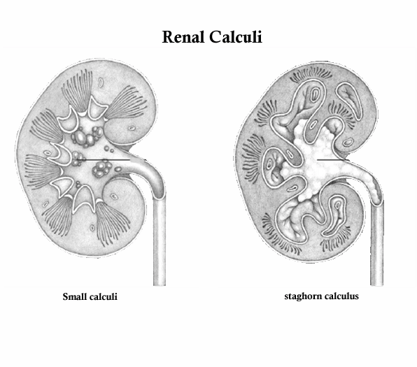
Renal Calculi/Kidney stones. Renal calculi, or nephrolithiasis, Kidney stones, are stones that form in the kidneys from the crystallization of minerals and other substances that normally dissolve in the urine. Renal calculi vary in size, with 90% less than 5 mm in diameter; some, however, grow large enough to prevent the natural passage of urine through the ureter. Renal calculi can form anywhere in the urinary tract, but they most commonly develop in the renal pelvis or calyces. Calculi may be solitary or multiple. Usualy these stones are composed of calcium salts. Other types are the struvite stones (which contain magnesium, ammonium, and phosphate), uric acid stones, and cystine stones. If the calculi remain in the renal pelvis or enter the ureter, they can damage renal parenchyma (functional tissue). Larger calculi can cause pressure necrosis. In certain locations, calculi cause obstruction, lead to hydronephrosis, and tend to recur. Renal calculi more common in males than females and are rare in blacks and children 

Causes for Renal Calculi/Kidney Stones
The precise cause of renal calculi is unknown, although Renal Calculi/Kidney stones are associated with dehydration, urinary obstruction, calcium levels, and other factors. 

  • Metabolic conditions such as renal tubular acidosis, elevated serum uric acid levels Hyperparathyroidism, renal tubular acidosis, elevated uric acid, defective metabolism of oxalate, a genetically caused defect in metabolism of cystine, and excessive intake of vitamin D or dietary calcium may predispose a person to renal calculi. 
  • Urinary tract infections associated with alkaline urine have been linked with calculus formation. Cystine stones are associated with hereditary renal disease 
  • Dehydration. Decreased water excretion concentrates calculus-forming substances. Patients who are dehydrated have decreased urine, with heavy concentrations of calculus-forming substances. Urinary obstruction leads to urinary stasis, a condition that contributes to calculus formation. 
  • Infection. Infected, scarred tissue may be a site for calculus development. In addition, infected calculi (usually magnesium ammonium phosphate or staghorn calculi) may develop if bacteria serve as the nucleus in calculus formation. Struvite calculus formation commonly results from Proteus infections, which may lead to destruction of renal parenchyma. 
  • Changes in urine pH. Consistently acidic or alkaline urine may provide a favorable medium for calculus formation, especially for magnesium ammonium phosphate or calcium phosphate calculi. 
  • Obstruction. Urinary stasis allows calculus constituents to collect and adhere, forming calculi. Obstruction also encourages infection, which compounds the obstruction. 
  • Immobilization. Immobility from spinal cord injury or other disorders allows calcium to be released into the circulation and, eventually, to be filtered by the kidneys. 
  • Renal Calculi/Kidney stones occur more often in men than in women 


Complications for Renal Calculi/Kidney stones
Calculi either remain in the renal pelvis and damage or destroy renal parenchyma, or they enter the ureter; large calculi in the kidneys cause pressure necrosis. Calculi in some sites cause obstruction, with resultant hydronephrosis, and tend to recur. Intractable pain and serious bleeding also can result from calculi and the damage they cause. 
Possible Complications for Renal Calculi/Kidney stones: 

  • Decrease or loss of kidney function 
  • Scarring, Kidney damage, 
  • Obstruction of the ureter (acute unilateral obstructive uropathy) 
  • stones Recurrence 
  • Urinary tract infection (UTI) 
  • renal colic 


Nursing Diagnosis for Renal Calculi/Kidney stones 
Nursing Diagnosis nursing care plans for Renal Calculi/Kidney stones determine by what we found in nursing assessment. Nursing Assessment nursing care plans for Renal Calculi/Kidney stones Typically, assessment findings vary depends with the size, location, and cause of the calculi: 
Patient history reveals a history of pain, and determine the intensity, duration, and location of the pain. The location of the pain varies according to the placement of the stone. The pain usually begins in the flank area but later may radiate into the lower abdomen and the groin. Ask if the pain had a sudden onset. Patients may relate a recent history of hematuria, nausea, vomiting, and anorexia. In cases in which a urinary tract infection is also present, the patient may report chills and fever. Determine the patient’s history to identify risk factors 
Physical Examination. Common symptom of renal calculi is severe pain, also referred as renal colic, which usually results from obstruction of large, rough calculi occlude the opening to the ureteropelvic junction and increase the frequency and force of peristaltic contractions. Pain intensity fluctuates and may be excruciating at its peak. 
Patient with calculi in the renal pelvis and calyces may complain of more constant, dull pain. He may also report pain and severe abdominal pain. The patient with severe pain also typically complains of nausea, vomiting and, possibly, fever and chills. Hematuria occur when calculi abrade a ureter, abdominal distention and, rarely, anuria 
Inspection reveals a patient in intense pain who is unable to maintain a comfortable position. Assess the patient for bladder distension. Monitor the patient for signs of an infection such as fever, chills, and increased white blood cell counts. Assess the urine for hematuria. Auscultate the patient’s abdomen for normal bowel sounds. Palpate the patient’s flank area for tenderness. Percussion of the abdominal area is normal, but percussion of the costovertebral angle elicits severe pain. 
Psychosocial Assessment: Patients with renal calculi may be extremely anxious because of the sudden onset of severe pain of unknown origin. Assess the patient’s ability to cope. Since diet and lifestyle may contribute to the formation of calculi, the patient may face lifestyle changes. Assess the patient’s ability to handle such changes. 

Diagnostic tests
Kidney-ureter-bladder (KUB) radiography, and Excretory urography, retrograde pyelography, Abdominal computed tomography scan, or Abdominal or kidney magnetic resonance imaging Kidney ultrasonography. Urine culture of a midstream Evaluated A 24-hour urine 
Other diagnostic test: Serial blood calcium and phosphorus levels indicate hyperparathyroidism and show an increased calcium level in proportion to normal serum protein levels. Blood protein levels are used to determine the level of free calcium unbound to protein. Increased blood uric acid levels may indicate gout. 

Nursing diagnosis Nursing Care Plans for Renal Calculi/Kidney stones
Common nursing diagnosis found in Nursing Care Plans for Renal Calculi/Kidney stones
Acute pain
Altered urinary elimination
Deficient knowledge (treatment plan)
Impaired urinary elimination
Ineffective tissue perfusion: Renal
Risk for imbalanced fluid volume
Risk for infection Risk for injury

Sample Nursing Care Plans for Renal Calculi/Kidney stones
Nursing diagnosis
Nursing outcome
Nursing Interventions
Evaluation
Acute pain related to inflammation, obstruction, and abrasion of urinary tract by migration of stones

·        Verbalizes reduced pain level

·        Administer prescribed  analgesic
·        Encourage patient to assume position that brings some relief.
·        Reassess pain frequently using pain scale.
·        Administer antiemetic  as indicated for nausea
·        Position the patient for comfort

Altered urinary elimination


Urinary Elimination:
Ability of the urinary system to filter wastes, conserve solutes, and
collect and discharge urine in a healthy pattern

Urinary Continence: Control of the elimination of urine
Self-Care: Toileting: Ability to toilet self
Assess degree of interference/disability
·       Determine client’s previous pattern of elimination and compare with current situation.
·                Frequency,
·                Urgency,
·                Burning,
·                Incontinence,
·                Nocturia/enuresis,
·                Size and force of urinary stream.
·       Provides information about degree of interference with elimination or may indicate bladder infection

·       Palpate bladder to assess retention. Fullness over bladder following voiding is indicative of inadequate emptying/retention and requires intervention.
·       Investigate pain which may be indicative of infection:
·                location,
·                duration,
·                 intensity;
·                Presence of bladder spasms, back or flank pain, etc.,.
·      Determine client’s usual daily fluid intake
Verbalize understanding of condition.
Identify causative factors. (Refer to specific NDs for incontinence/retention as appropriate.)
Achieve normal elimination pattern or participate in measures to correct/compensate for
defects.
Demonstrate behaviors/techniques to prevent urinary infection.
Manage care of urinary catheter, or stoma and appliance following urinary diversion.

Saturday, February 20, 2010

The diagnosis of delusional disorder can be made when a person exhibits non-bizarre delusions of at least 1 month duration that cannot be attributed to other psychiatric disorders. Non-bizarre delusions must be about phenomena that, although not real, are within the realm of being possible. In general, the patient’s delusions are well systematized and have been logically developed. The person’s behavioral and emotional responses to the delusions appear to be appropriate. Usually the person’s functioning and personality are well preserved and show minimal deterioration if at all. 
Characteristics of delusional disorder

  • Non-bizarre delusions of at least 1 month’s duration 
  • No positive or negative symptoms of schizophrenia present 
  • Tactile or olfactory hallucinations may be present and related to the delusional theme 
  • Functioning not markedly impaired and behavior not obviously bizarre or odd 
  • Only brief mood episodes, if any 
  • Not due to direct physiologic effects of a chemical or a general medical condition 


Etiology of delusional disorder 
Etiology of the delusional disorder is unknown. Risk factors associated with the disorder include advanced age, sensory impairment/isolation, family history, social isolation, personality features (e.g. unusual interpersonal sensitivity), and recent immigration. Some have reported higher association of delusional disorder with widowhood, celibacy, and history of substance abuse. Age of onset is later than schizophrenia and earlier in men compared to women. 

Subtypes of delusional disorder 

Persecutory Type 
Here the person affected believes that he or she is being followed, spied on, poisoned or drugged, harassed, or conspired against. The person affected may get preoccupied by small slights that can become incorporated into the delusional system. These individuals may resort to legal actions to remedy perceived injustice. Individuals suffering from these delusions often become resentful and angry with a potential to get violent against those believed to be against them. 

Jealous Type 
Individuals with this subtype have the delusional belief that their spouses/lovers are unfaithful. Jealousy is a powerful emotion and when it occurs in delusional disorder or as part of another condition, it can be potentially dangerous and has been associated with violence including suicidal and homicidal behavior. Delusions of infi delity have also been called conjugal paranoia . The term Othello syndrome has been used to describe morbid jealousy. 

Erotomanic Type 
Persons with delusional disorder of the erotomanic type have delusions of being loved by another. The patient believes that a perceived suitor, usually more socially prominent than herself, is in love with her. Erotomania shares many features with, is derived from, and is often referred to as de Clerambault’s syndrome. 

Somatic Type 
Delusional disorder with somatic delusions has been called monosymptomatic hypochondriacal psychosis . This disorder differs from other conditions with hypochondriacal symptoms in degree of reality impairment. Munro ( 1991 ) has described the largest series of cases and has used content of delusions to defi ne three main types: 
Delusions of Infestations (Including Parasitosis). 
Delusional parasitosis has been described in association with many physical illnesses such as vitamin B 12 defi ciency, pellagra, neurosyphilis, multiple sclerosis, thalamic dysfunction, hypophyseal tumors, diabetes mellitus, severe renal disease, hepatitis, hypothyroidism, mediastinal lymphoma, and leprosy. Use of cocaine and presence of dementia has also been reported. Psychogenic parasitosis was also known as Ekbom’s syndrome before being referred to as delusional parasitosis . 
Delusions of Dysmorphophobia 
This condition includes delusions such as of misshapenness, personal ugliness, or exaggerated size of body parts. 
Delusions of Foul Body Odors or Halitosis. 
This is also called olfactory reference syndrome. 

Grandiose Type 
This is also referred to as megalomania . In this subtype, the central theme of the delusion is the grandiosity of having made some important discovery or having great talent. Sometimes there may be a religious theme to the delusional thinking such that the person believes that he or she has a special message from god. 

Mixed Type 
This subtype is reserved for those with two or more delusional themes. However, it should be used only where it is difficult to clearly discern one theme of delusion. This subtype is used for cases in which the predominant delusion cannot be subtyped within the above mentioned categories. A possible example is certain delusions of misidentification, for example, Capgras’s syndrome , named after the French psychiatrist who described the ‘illusions of doubles.’ The delusion here is the belief that a familiar person has been replaced by an imposter. A variant of this is Fregoli’s syndrome where the delusion is that the persecutors or familiar persons can assume the guise of strangers and the very rare delusion that familiar persons could change themselves into other persons at will (intermetamorphosis). 

COMPLICATIONS of delusional disorder If left undiagnosed, untreated, or ineffectively treated, schizophrenia can lead to profound inability to function and contribute to the problem of homelessness in our society. Neglect of other medical conditions; therefore, complications due to untreated medical illness are common. Depression and suicide. Substance use, abuse, or dependency. 

Nursing Diagnosis Delusional Disorder 
Nursing Diagnosis for Delusional Disorder determine from what we found in Nursing Assessment Nursing Care Plans for Delusional Disorder. 

Nursing assessment nursing care Plans for Delusional Disorders Assess for positive symptoms of schizophrenia. These symptoms reflect aberrant mental activity and are usually present early in the first phase of the schizophrenic illness. 
Alterations in Thinking 

  • Delusion: false, fixed belief that is not amenable to change by reasoning. The most frequent elicited delusions include: Ideas of reference. Delusions of grandeur. Delusions of jealousy. Delusions of persecution. Somatic delusions. 
  • Loose associations: the thought process becomes illogical and confused. 
  • Neologisms: made-up words that have a special meaning to the delusional person. 
  • Concrete thinking: an overemphasis on small or specific details and an impaired ability to abstract. 
  • Echolalia: pathologic repeating of another’s words. 
  • Clang associations: the meaningless rhyming of a word in a forceful way. 
  • Word salad: a mixture of words that is meaningless to the listener. 


Alterations in Behavioral Responses 

  • Bizarre behavioral patterns Motor agitation and restlessness Automatic obedience or robotlike movement Autonomic obedience or robotlike movement Negativism Stereotyped behaviors Stupor Waxy flexibility (allowing another person to reposition extremities) 
  • Agitated or impulsive behavior 
  • Assess for negative symptoms of schizophrenia that reflect a deficiency of mental functioning Alogia (lack of speech) Anergia ( inability to react) Anhedonia ( inability to experience pleasure) Avolition (lack of motivation or initiation) Poor social functioning Poverty of speech Social withdrawal Thought blocking 
  • Assess for associated symptoms of schizophrenia Substance use, abuse, or dependence Depression Fantasy Violent or aggressive behavior Water intoxication Withdrawal 


Common nursing diagnosis found in Nursing Care Plans for Delusional Disorder 

  • Disturbed Thought Processes related to perceptual and cognitive distortions, as demonstrated by suspiciousness, defensive behavior, and disruptions in thought
  • Social Isolation related to an inability to trust 
  • Activity Intolerance related to adverse reactions to psychopharmacologic drugs 
  • Ineffective Coping related to misinterpretation of environment and impaired communication ability 
  • Risk for Self-directed or Other-directed Violence related to delusional thinking and hallucinatory experiences 


Nursing Care Plans for Delusional Disorder 
Nursing Care Plans For Delusional Disorder, delusional disorder diagnosis can be made when a person exhibits nonbizarre delusions of at least 1 month duration that cannot be attributed to other psychiatric disorders. Nonbizarre delusions must be about phenomena that, although not real, are within the realm of being possible. In general, the patient’s delusions are well systematized and have been logically developed. The person’s behavioral and emotional responses to the delusions appear to be appropriate. Usually the person’s functioning and personality are well preserved and show minimal deterioration if at all. 

Nursing Care Plans Delusional Disorder with nursing diagnosis; Disturbed Thought Processes, Social Isolation, Activity Intolerance, Ineffective Coping, Risk for Self-directed or Other-directed Violence.
NURSING DIAGNOSE
NURSING OUTCOME
INTERVENTION
EVALUATION
Disturbed Thought Processes related to perceptual and cognitive distortions, as demonstrated by suspiciousness, defensive behavior, and disruptions in thought

Patient showed the Differentiation Between Delusions and Reality 
  • Provide patient with honest and consistent feedback in a nonthreatening manner.
  • Avoid challenging the content of patient's behaviors.
  • Focus interactions on patient's behaviors.
  • Administer drugs as prescribed while monitoring and documenting patient's response to the drug regimen.
  • Use simple and clear language when speaking with patient.
  • Explain all procedures, tests, and activities to patient before starting them, and provide written or video material for learning purposes.

Exhibits improved reality orientation, concentration, and attention span as demonstrated through speech and behavior
Social Isolation related to an inability to trust

Patient showed the Promoting Socialization
  • Encourage patient to talk about feelings in the context of a trusting, supportive relationship.
  • Allow patient time to reveal delusions to you without engaging in a power struggle over the content or the reality of the delusions.
  • Use a supportive, empathic approach to focus on patient's feelings about troubling events or conflicts.
  • Provide opportunities for socialization and encourage participation in group activities.
  • Be aware of patient's personal space and use touch judiciously.
  • Help patient to identify behaviors that alienate significant others and family members.

Communicates with family and staff in a clear manner without evidence of loose, dissociated thinking

Activity Intolerance related to adverse reactions to psychopharmacologic drugs

Patient showed the Improving Activity Tolerance
  • Assess patient's response to prescribed antipsychotic drug.
  • Collaborate with patient and occupational and physical therapy specialists to assess patient's ability to perform ADLs.
  • Collaborate with patient to establish a daily, achievable routine within physical limitations.
  • Teach strategies to manage adverse effects of antipsychotic drug that affect patient's functional status, including:
    • Change positions slowly
    • Gradually increase physical activities
    • Limit overdoing it in hot, sunny weather
    • Use sun precautions
    • Use caution in activities if extrapyramidal symptoms develop.

Independently maintains personal hygiene without fatigue
Ineffective Coping related to misinterpretation of environment and impaired communication ability

Patient showed the Improving Coping with Thoughts and Feelings

  • Encourage patient to express feelings.
  • Focus on patient's feelings and behavior.
  • Provide honest perceptions of reality and feedback about symptoms and behaviors.
  • Encourage patient to explore adaptive behaviors that increase abilities and success in socializing and accomplishing ADLs.
  • Decrease environmental stimuli.

Attends group activities

Risk for Self-directed or Other-directed Violence related to delusional thinking and hallucinatory experiences

Safety appears
  • Monitor patient for behaviors that indicate increased anxiety and agitation.
  • Collaborate with patient to identify anxious behaviors as well as the causes.
  • Tell patient that you will help with maintaining behavioral control.
  • Establish consistent limits on patient's behaviors and clearly communicate these limits to patient, family members, and health care providers.
  • Secure all potential weapons and articles from patient's room and the unit environment that could be used to inflict an injury.
  • To prepare for possible continued escalation, form a psychiatric emergency assist team and designate a leader to facilitate an effective and safe aggression-management process.
  • Determine the need for external control, including seclusion or restraints. Communicate the decision to patient and put plan into action.
  • Frequently monitor patient within the guidelines of facility's policy on restrictive devices and assess the patient's level of agitation.
  • When patient's level of agitation begins to decrease and self-control is regained, establish a behavioral agreement that identifies specific behaviors that indicate self-control against a reescalation of agitation.

Remains free from harm or violent acts