Friday, June 18, 2010

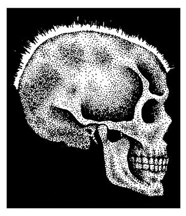
skull abnormality in a patient with thalassemia major
Thalassemia is a group of hereditary hemolytic anemia, which Marked by a deficit in the production of hemoglobin. Where the damage occurred in the red blood cells in the blood vessel so that the age erythrocyte be short. It is characterized by defective synthesis in one or more of the polypeptide chains necessary for hemoglobin production. Because Thalassemia affects hemoglobin production, it also impairs red blood cell (RBC) synthesis. 
There are two main types of Thalassemia: ”alpha” and “beta.” The names refer to the part of the hemoglobin molecule that is missing or mutated. 
Two pairs of polypeptide chains”alpha and beta chains” make up hemoglobin. In Thalassemia, diminished synthesis can affect either pair. Structurally, the chains are normal, but the genetic defect decreases their number. In alpha Thalassemia, alpha chain synthesis slows; in beta-Thalassemia, beta chain synthesis slows. Some patients with beta-Thalassemia have no normal hemoglobin only hemoglobin S and the minor hemoglobin’s. 

Causes for Thalassemia 
Thalassemia major Causes by homozygous inheritance of the partially dominant autosomal gene Thalassemia intermedia Causes by homozygous inheritance of the partially dominant autosomal gene. Thalassemia minor is caused by heterozygous inheritance of the same gene. In All type of Thalassemia deficiency of beta polypeptide chain production impairs hemoglobin synthesis and results in continual production of fetal hemoglobin, even after the neonatal period. 

Complications for Thalassemia 
Pathologic fractures Cardiac arrhythmias Heart failure Other complications that result from iron deposits in the heart and other tissues caused by repeated blood transfusions. 

Treatment for Thalassemia 
Patients with Thalassemia minor usually do not require any specific treatment. Treatment for patients with Thalassemia major includes chronic blood transfusion therapy, iron chelation, splenectomy, and allogeneic hematopoietic transplantation. References: http://en.wikipedia.org/wiki/Thalassemia 

Nursing Assessment
Thalassemia major: severe anemia, splenomegaly or hepatomegaly with abdominal enlargement, frequent infections, bleeding tendencies e.g. epistaxis, and anorexia, small bodies and large heads, mentally disabled, mongoloid. Thalassemia intermediate: anemia, jaundice, and splenomegaly, hemosiderosis caused by increased intestinal absorption of iron. Thalassemia minor: mild anemia usually with no signs or symptoms. 

Diagnostic Test for Thalassemia 
RBC count and hemoglobin level X-rays of the skull and long bones 

Nursing Diagnosis
Activity intolerance Deficient knowledge (treatment regimen) Delayed growth and development Disturbed body image Ineffective tissue perfusion: Cardiopulmonary Interrupted family processes Risk for infection 

Nursing care Plans for Thalassemia 
Treatment of thalassemia major is essentially supportive. For example, infections require prompt treatment with the appropriate antibiotic. Transfusions of packed RBCs raise the hemoglobin level but must be used judiciously to minimize iron overload. Thalassemia intermedia and thalassemia minor generally don’t require treatment. Iron supplements are contraindicated in all forms of thalassemia. Treatment of children is more difficult. Regular blood transfusions may minimize physical and mental retardation, but transfusions increase the risk of deadly hemosiderosis and iron overload. Continuous subcutaneous infusion of an iron-chelating agent may help produce a negative overall iron balance. If rapid splenic sequestration of transfused RBCs necessitates more transfusions, a splenectomy may be performed. 

Nursing outcomes 
Patient and parents will: Verbalize the importance of balancing activity, as tolerated, with frequent rest periods. Demonstrate age-appropriate skills and behaviors to the extent possible. Express positive feelings about himself. Develop no cardiac arrhythmias. Voice feelings and concerns related to the patient’s illness. Remain free from signs and symptoms of infection Nursing interventions nursing care plans for Thalassemia Watch for adverse reactions during and after RBC transfusions. Collaborative an antibiotic, and observe the patient for adverse reactions. Provide an adequate diet, Encourage the patient to drink plenty of fluids. Provide emotional support Help the patient and his family cope for chronic nature of Explain the need for lifelong transfusions. 

Patient teaching
Explain how to prevent infection e.g. nutrition, wound care Tell about signs of hepatitis and iron overload, which are always possible with frequent transfusions. Explain why child must avoid strenuous athletic activity to avoid pathologic fractures.

Friday, June 11, 2010

Acute Pain
NANDA Nursing Diagnosis for Acute Pain Related factors R/T trauma, injuring agents (biological, chemical, physical, psychological) 
Suggestion on using NANDA Nursing Diagnosis Acute Pain: Distinguish between acute pain Nursing diagnosis with the chronicles pain Nursing diagnosis. One of the two sets is that diagnosis of a painful time. ONSET acute pain is less than 6 months, while the painful chronicles ONSET it is more than 6 months. If you only have two diagnoses to indicate, pain is acute pain and chronic pain. Thus, there is no automatic diagnosis Crashes feel comfortable or feel comfortable painful chronic pain. 
Definition Nursing Diagnosis for Acute Pain: Unpleasant sensory and emotional experience arising from actual or potential tissue damage or described in terms of such damage (International Association for the Study of Pain); sudden or slow onset of any intensity from mild to severe with an anticipated or predictable end and a duration of less than 6 months 

Nursing Outcomes Client Will: Report pain is relieved / controlled. Follow prescribed pharmacological regimen. Verbalize methods that provide relief. Demonstrate use of relaxation skills and diversion activities as indicated for individual situation. 

Nursing Priority Nursing Diagnosis for Acute Pain To assess etiology/precipitating contributory factors: evaluate client’s response to pain: assist client to explore methods for alleviation/control of pain 

Sample Clinical Applications using Nursing Diagnosis for Acute Pain: Traumatic injuries, surgical procedures, infections, cancer, burns, skin lesions, gangrene, thrombophlebitis/pulmonary embolus, neuralgia

Thursday, June 10, 2010

Malignant Brain Tumors
Primary brain tumors comprise a diverse group of neoplasm’s that are often malignant and refractory to treatment, malignant brain tumors. More common in males than in females, malignant brain tumors (gliomas, meningiomas, and schwannomas) they cause central nervous system (CNS) changes by invading and destroying tissues and by secondary effects mainly compression of the brain, cranial nerves, and cerebral vessels; cerebral edema; and increased intracranial pressure ICP. Tumors can occur at any age. In adults, incidence is highest between ages 40 and 60, and the most common tumor types are gliomas and meningiomas. They usually occur above the covering of the cerebellum (supratentorial tumors). Most tumors in children occur before age one or between ages 2 and 12. The most common are astrocytomas, medulloblastomas, ependymomas, and brain stem gliomas. Brain tumors are one of the most common causes of cancer death in children. 

Primary brain tumors classified by their siteof origin 
  • CNS tumors that originate in glial tissue Gliomas, occurring in the glial, or supportive tissues around the brain: Astrocytomas Brain-stem gliomas Ependymomas Oligodendrogliomas 
  • CNS tumors that do not originate in glial tissue: Medulloblastoma Craniopharyngiomas Germinomas Pineal-region tumors 


Causes of Brain Tumor 
Brain tumors may be attributed to a familial link or to exposure to ionized radiation (a known environmental risk). Brain tumors may result from metastasis in 20% to 40% of patients with other cancers. In some cases, the cause remains unknown. 

Risk Factors of Primary Brain Tumors 
Hereditary syndromes (proven): tuberous sclerosis, neurofibromatosis types 1 and 2, nevoid basal cell carcinoma syndrome, Turcot’s syndrome, and Li-Fraumeni syndrome Family History of brain tumors History of prior cancer Exposure to infectious agents Allergies (possible reduced risk) Head trauma Drugs and medications Dietary history: N-nitroso compounds, oxidants, antioxidants Tobacco usage Alcohol consumption Ionizing radiation exposure Cellular telephones, Power frequency electromagnetic field exposure 

Complications 
In malignant brain tumors, life-threatening complications from increasing ICP include coma, respiratory or cardiac arrest, and brain herniation

Monday, June 7, 2010

Cervical cancer is the third most common cancer of the female reproductive system. Cancer of the cervix is one type of primary uterine cancer (the other being uterine-endometrial cancer) and is predominately epidermoid. Invasive cervical cancer is the third most common female pelvic cancer. The death rate from cervical cancer has steadily declined over the past 50 years owing to the increased use of the Papanicolaou exam, which detects cervical changes before cancer develops. Three types of cervical cancer are: Dysplasia, Carcinoma in situ (CIS) and Invasive carcinoma 
Preinvasive cancer ranges from minimal cervical dysplasia, in which the lower third of the epithelium contains abnormal cells, to carcinoma in situ, in which the full thickness of epithelium contains abnormally proliferating cells (also known as cervical intraepithelial neoplasia). Preinvasive cancer is curable in 75% to 90% of patients with early detection and proper treatment. If untreated, it may progress to invasive cervical cancer, depending on the form. 
CIS is carcinoma confined to the epithelium. The full thickness of the epithelium contains abnormally proliferating cells. Both dysplasia and CIS are considered preinvasive cancers and, with early detection, have a 5-year survival rate of 73% to 92%. 
In invasive disease, cancer cells penetrate the basement membrane and can spread directly to contiguous pelvic structures or disseminate to distant sites by way of lymphatic routes. In 95% of cases, the histologic type is squamous cell carcinoma, which varies from well-differentiated cells to highly anaplastic spindle cells. Only 5% of cases are adenocarcinomas. Invasive cancer typically occurs between ages 30 and 50; it rarely occurs younger than age 20. 

Cervical cancer stage (source: http://en.wikipedia.org) 
Cervical cancer is staged by the International Federation of Gynecology and Obstetrics (FIGO) staging system, which is based on clinical examination, rather than surgical findings. It allows only the following diagnostic tests to be used in determining the stage: palpation, inspection, colposcopy, endocervical curettage, hysteroscopy, cystoscopy, proctoscopy, intravenous urography, and X-ray examination of the lungs and skeleton, and cervical conization. 
The TNM staging system for cervical cancer is analogous to the FIGO stage. 

Stage 0 – full-thickness involvement of the epithelium without invasion into the stroma (carcinoma in situ) 
Stage I – limited to the cervix 
IA – diagnosed only by microscopy; no visible lesions 
          IA1 – stromal invasion less than 3 mm in depth and 7 mm or less in horizontal spread 
          IA2 – stromal invasion between 3 and 5 mm with horizontal spread of 7 mm or less 
IB – visible lesion or a microscopic lesion with more than 5 mm of depth or horizontal spread of more than 7 mm 
          IB1 – visible lesion 4 cm or less in greatest dimension 
          IB2 – visible lesion more than 4 cm 
Stage II – invades beyond cervix
          IIA – without parametrial invasion, but involve upper 2/3 of vagina
          IIB – with parametrial invasion 
Stage III – extends to pelvic wall or lower third of the vagina
          IIIA – involves lower third of vagina
          IIIB – extends to pelvic wall and/or causes hydronephrosis or non-functioning kidney 
IVA – invades mucosa of bladder or rectum and/or extends beyond true pelvis 
IVB – distant metastasis 

Causes for Cervical cancer 
Worldwide studies suggest that Causes for Cervical cancer is sexually transmitted human papillomaviruses (HPVs). Certain strains of HPV (16, 18, and 31) are associated with an increased risk of cervical cancer. Several predisposing factors have been related to the development of cervical cancer: intercourse at a young age), multiple sexual partners, and herpesvirus 2 and other bacterial or viral venereal infections. Genetic considerations While most risk factors for cervical cancer are environmental, some studies have found that the daughters or sisters of cervical cancer patients are more likely to get the disease. 

Complications of Cervical cancer 
Disease progression can cause flank pain from sciatic nerve or pelvic wall invasion and hematuria and renal failure associated with bladder involvement.

  • Ureteric obstruction 
  • Intermenstrual PV bleed 
  • Vesicovaginal fistula 
  • Post-menopausal PV bleed 
  • Uterine enlargement 
  • Menorrhagia 

Nursing Assessment
Patient’s history, early cervical cancer usually asymptomatic, establishes a thorough history with particular attention to the presence of the risk factors and the woman’s menstrual history. assess a history of later symptoms of cervical cancer, including abnormal bleeding or spotting between periods or after menopause, metrorrhagia or menorrhagia, dysparuenia and postcoital bleeding; leukorrhea in increasing amounts and changing over time from watery to dark and foul; and a history of chronic cervical infections. Determine if the patient has experienced weight gain or loss; abdominal or pelvic pain, often unilateral, radiating to the buttocks and legs, or other symptoms associated with neoplasms, such as fatigue. The patient history includes abnormal vaginal bleeding, such as a persistent vaginal discharge that may be yellowish, blood-tinged, and foul-smelling; postcoital pain and bleeding; and bleeding between menstrual periods or unusually heavy menstrual periods. The patient history may suggest one or more of the predisposing factors for this disease. 
Physical Examination. Pelvic examination. Observe the patient’s external genitalia for signs of inflammation, bleeding, discharge, or local skin or epithelial changes. Palpate for motion tenderness of the cervix (Chandelier’s sign); a positive Chandelier’s sign (pain on movement) usually indicates an infection. Also examine the size, consistency (hardness may reflect invasion by neoplasm), shape, mobility (cervix should be freely movable), tenderness, and presence of masses of the uterus and adnexa. If the cancer has advanced into the pelvic wall, the patient may report gradually increasing flank pain, which can indicate sciatic nerve involvement. Leakage of urine may point to metastasis into the bladder with formation of a fistula. Leakage of stool may indicate metastasis to the rectum with fistula development. 

Diagnostic test
Papanicolaou examination ((Pap smear) Colposcopy followed by punch biopsy or cone biopsy The Vira/Pap test to examination of the specimen’s deoxyribonucleic acid (DNA) structure to detect HPV 

Nursing diagnosis
Common nursing diagnosis found in nursing care plans for Cervical Cancer:

  • Pain (acute) related to postprocedure swelling and nerve damage 
  • Anxiety 
  • Fear 
  • Impaired physical mobility 
  • Impaired skin integrity 
  • Ineffective coping 
  • Ineffective sexuality patterns 
  • Risk for infection Sexual dysfunction 

Cervical cancer is the third most common cancer of the female reproductive system. Cancer of the cervix is one type of primary uterine cancer (the other being uterine-endometrial cancer) and is predominately epidermoid. Invasive cervical cancer is the third most common female pelvic cancer. The death rate from cervical cancer has steadily declined over the past 50 years owing to the increased use of the Papanicolaou exam, which detects cervical changes before cancer develops.

Nursing Key outcomes
Pain control; Pain: Disruptive effects; Well-being, after nursing interventions patient will Report feeling less pain. Report feelings of reduced anxiety. Verbalize her concerns and fears related to her diagnosis and condition. Maintain joint mobility and range of motion. Free from breakdown. Demonstrate adaptive coping behaviors. Resume normal sexual activity patterns to the fullest extent possible. Remain free from signs or symptoms of infection. The patient and partner will express feelings and perceptions about changes in sexual performance.

Nursing interventions nursing care plans for Cervical Cancer 
Analgesic administration; Pain management; Meditation; Transcutaneous electric nerve stimulation (TENS); Hypnosis; Heat/cold application
Collaborative 
If you assist with a biopsy, drape and prepare the patient as for a routine Pap test and pelvic examination. Have a container of formaldehyde ready to preserve the specimen during transfer to the pathology laboratory. Assist the physician as needed, and provide support for the patient throughout the procedure. If you assist with cryosurgery or laser therapy, drape and prepare the patient as for a routine Pap test and pelvic examination. Assist the physician as necessary, and provide support for the patient throughout the procedure. Preinvasive lesions (CIS) can be treated by conization, cryosurgery, laser surgery, or simple hysterectomy (if the patient’s reproductive capacity is not an issue). All conservative treatments require frequent follow-up by Pap tests and colposcopy because a greater level of risk is always present for the woman who has had CIS Administer analgesics and prophylactic antibiotics, as ordered.
Independent 
Listen to the patient’s fears and concerns, and offer reassurance when appropriate. Encourage her to use relaxation techniques to promote comfort during diagnostic procedures. When a patient requires surgery, prepare her mentally and physically for the surgery and the postoperative period. After any surgery, monitor vital signs every 4 hours. Watch for and immediately report signs of complications, such as bleeding, abdominal distention, severe pain, and wheezing or other breathing difficulties. Encourage deep breathing and coughing. Check to see whether the radioactive source is to be inserted while the patient is in the operating room (preloaded) or at bedside (afterloaded). If the source is preloaded, the patient returns to her room hot and safety precautions begin immediately. Remember that safety precaution time, distance, and shielding begin as soon as the radioactive source is in place. Inform the patient that she will require a private room. Check the patient’s vital signs every 4 hours Assist the patient with range-of-motion arm exercises. Avoid leg exercises and other body movements that could dislodge the source. If ordered, administer a tranquilizer to help the patient relax. Provide activities that require minimal movement. Watch for treatment complications by listening to and observing the patient and monitoring laboratory studies and vital signs. When appropriate, perform measures to prevent or alleviate complications.

Patient teaching, discharge and home healthcare guidelines for patients with Cervical Cancer: 
Be sure the patient and family understand any pain medication prescribed, including dosage, route, action, and side effects. Reassure the patient that this disease and Cervical Cancer care treatment should not radically alter her lifestyle or prohibit sexual intimacy. Tell to the patient all the post procedure complications. Ensure that the patient understands the need for ongoing Pap smears if appropriate. Vaginal cytological studies are recommended at 4-month intervals for 2 years, every 6 months for 3 years, and then annually. Explain the importance of complying with follow-up visits to the gynecologist and oncologist. Stress the value of these visits in detecting disease progression or recurrence

Biopsy
Explain to the patient that she may feel pressure, minor abdominal cramps, or a pinch from the punch forceps. Reassure her that the pain will be minimal because the cervix has few nerve endings.

Cryosurgery
Explain to the patient that the procedure takes about 15 minutes, during which time the physician uses refrigerant to freeze the cervix. Caution to the patients that she may experience abdominal cramps, headache, and sweating, but reassure her that she will feel little, if any, pain.

Laser surgery
Explain to the patient the laser surgery procedure takes about 30 minutes and may cause abdominal cramps. After excision biopsy, cryosurgery, or laser therapy, tell the patient to expect a discharge or spotting for about 1 week. Advise her not to douche, use tampons, or engage in sexual intercourse during this time. Caution her to report signs of infection. Stress the need for a follow-up Pap test and a pelvic examination in 3 to 4 months and periodically thereafter. Also, tell her what to expect postoperatively if a hysterectomy is necessary.

Preloaded internal radiation therapy
Tell to the patient that preloaded internal radiation therapy procedure requires hospital stay, bowel preparation, a povidoneiodine vaginal douche, a clear liquid diet, and nothing by mouth the night before the implantation. It also requires an indwelling urinary catheter. Inform the patient that preloaded internal radiation therapy is performed in the operating room under general anesthesia.

After loaded internal radiation therapy
Explain to the patient that a member of the radiation team implants the source after the patient returns to her room from surgery. Remind the patient to watch for and report uncomfortable adverse effects, warn the patient to avoid people with obvious infections during therapy. Inform the patient that vaginal narrowing caused by scar tissue can occur after internal radiation. Describe the complications that can occur after high-dose radiation therapy.

Tuesday, May 25, 2010

Abruptio Placentae (Placenta Abruption)
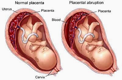
Abruptio placentae also called placental abruption occur when the placenta prematurely separates from the uterine wall, usually after the 20th week of gestation, producing hemorrhage. This disorder may be classified according to the degree of placental separation and the severity of maternal and fetal symptoms. It is characterized by a triad of symptoms: vaginal bleeding, uterine hypertonus, and fetal distress. It can occur during the prenatal or intrapartum period. Abruptio placentae is most common in multigravidas usually in women older than age 35 and is a common cause of bleeding during the second half of pregnancy. On heavy maternal bleeding generally necessitates termination of the pregnancy. The fetal prognosis depends on the gestational age and amount of blood lost. The maternal prognosis is good if hemorrhage can be controlled. 

Grading System for Abruptio Placentae (placenta abruption) 
  • Grade 0 Less than 10% of the total placental surface has detached; the patient has no symptoms; however, a small retroplacental clot is noted at birth. 
  • Grade I approximately 10%–20% of the total placental surface has detached; vaginal bleeding and mild uterine tenderness are noted; however, the mother and fetus are in no distress. 
  • Grade II Approximately 20%–50% of the total placental surface has detached; the patient has uterine tenderness and tetany; bleeding can be concealed or is obvious; signs of fetal distress are noted; the mother is not in hypovolemic shock. 
  • Grade III More than 50% of the placental surface has detached; uterine tetany is severe; bleeding can be concealed or is obvious; the mother is in shock and often experiencing coagulopathy; fetal death occurs. 

Central abruption
Central abruption, the separation occurs in the middle, and bleeding is trapped Between the detached placenta and the uterus, concealing the hemorrhage 

Marginal abruption
Marginal abruption, separation begins at the periphery and bleeding accumulates between The membranes and the uterus and eventually passes through the cervix, becoming an external hemorrhage. 

Causes for Abruptio Placentae (placenta abruption) 
The cause of abruptio placentae is unknown; however, any condition that causes vascular changes at the placental level may contribute to premature separation of the placenta. Predisposing factors include: Traumatic injury. Placental site bleeding from a needle puncture during amniocentesis, Chronic or pregnancy-induced hypertension. Multiparity Short umbilical cord Dietary deficiency Smoking Advanced maternal age Pressure on the vena cava from an enlarged uterus. 
The spontaneous rupture of blood vessels at the placental bed may result from a lack of resiliency or to abnormal changes in the uterine vasculature. The condition may be complicated by hypertension or by an enlarged uterus that can’t contract sufficiently to seal off the torn vessels. Consequently, bleeding continues unchecked, possibly shearing off the placenta partially or completely. 

Complications for Abruptio Placentae (placenta abruption) 
Hemorrhage and shock. Renal failure, Disseminated intravascular coagulation. Maternal and fetal death. 

Nursing Assessment
Abruptio placentae produce a wide range of clinical effects, depending on the extent of placental separation and the amount of blood lost from maternal circulation. Obtain patient history obstetric history. Determine the date of the last menstrual period to calculate the estimated day of delivery and gestational age of the infant. Inquire about alcohol, tobacco, and drug usage, and any trauma or abuse situations during pregnancy Mild Abruptio placentae with marginal separation usually report mild to moderate vaginal bleeding, vague lower abdominal discomfort, and mild to moderate abdominal tenderness. Moderate Abruptio placentae are about 50% placental separation usually report continuous abdominal pain and moderate, dark red vaginal bleeding. Onset of symptoms may be gradual or abrupt. Vital signs may indicate impending shock. Palpation reveals a tender uterus that remains firm between contractions. Severe Abruptio placentae about 70% placental separations patient usually report abrupt onset of agonizing, unremitting uterine pain (described as tearing or knifelike) and moderate vaginal bleeding. Vital signs indicate rapidly progressive shock. Palpation reveals a tender uterus with board like rigidity. Uterine size may increase in severe concealed abruptions. Psychosocial Assessment to understanding patient’s situation and also the significant other’s degree of anxiety, coping ability, and willingness to support the patient 

Diagnostic tests for Abruptio Placentae (placenta abruption) 
Pelvic examination under double setup Ultrasonography Decreased hemoglobin level Decreased platelet count. Periodic assays for fibrin split products aid in monitoring the progression of abruptio placentae and in detecting DIC. 

Treatment for Abruptio Placentae (placenta abruption) Medical Treatment management goals of abruptio placentae are to assess, control, and restore the amount of blood lost and to deliver a viable infant and prevent coagulation disorders. After determining the severity of placental abruption and appropriate fluid and blood replacement, prompt cesarean delivery is necessary if the fetus is in distress. If the fetus isn’t in distress, monitoring continues; delivery is usually performed at the first sign of fetal distress. 

Nursing diagnosis
Primary nursing diagnosis fluid volume deficit related to blood loss. Common nursing diagnosis fond in Nursing Care Plans for Abruptio Placentae (placenta abruption): Acute pain Anxiety Deficient fluid volume Dysfunctional grieving Fear Ineffective coping Ineffective tissue perfusion: Cardiopulmonary  

Key outcomes  the patient will: 
Express feelings of comfort. Express feelings of reduced anxiety. Communicate feelings about the situation. Discuss fears and concerns. Use available support systems, such as family and friends, to aid in coping. Remain hemodynamically stable. Patient’s fluid volume will remain within normal parameters. 

Nursing interventions
Monitor Vital sign; blood pressure, pulse rate, respirations, central venous pressure, intake and output, and amount of vaginal bleeding. Monitor fetal heart rate electronically. If vaginal delivery is elected, provide emotional support during labor. Because of the neonate’s prematurity, the mother may not receive an analgesic during labor and may experience intense pain. Reassure the patient of her progress through labor, and keep her informed of the fetus’s condition. Encourage the patient and her family to verbalize their feelings. Help them to develop effective coping strategies. Refer them for counseling, if necessary. 

Patient teaching discharge and home healthcare guidelines
Teach the patient to identify and report signs of placental abruption, such as bleeding and cramping. Explain procedures and treatments to allay patient’s anxiety. Teach the patient to notify the doctor and come to the hospital immediately if she experiences any bleeding or contractions. Prepare the patient and her family for the possibility of an emergency cesarean delivery, the delivery of a premature neonate, and the changes to expect in the postpartum period. Offer emotional support and an honest assessment of the situation. Tactfully discuss the possibility of neonatal death. Inform the patient that the neonate’s survival depends primarily on gestational age, the amount of blood lost, and associated hypertensive disorders. Inform the patient that frequent monitoring and prompt management greatly reduce the risk of death. 

After Postpartum Patient teaching discharge and home healthcare guidelines 
Give the usual postpartum instructions for avoiding complications. Inform the patient that she is at much higher risk of developing abruptio placentae in subsequent Pregnancies. Instruct the patient on how to provide safe care of the infant. Provide a list of referrals to the patient and significant others to help them manage their loss, If the fetus has not Survived

Monday, May 24, 2010

Bulimia Nervosa
Bulimia nervosa the binge and purge syndrome is an eating disorder, the essential features of bulimia nervosa include eating binges followed by feelings of guilt, humiliation, and self deprecation guilt, and anxiety over fear of weight gain. Characterized by extreme overeating, followed by self induced vomiting and abuse of laxatives, diuretics, strict dieting or fasting to overcome the effects of the binges. Unless the patient devotes an excessive amount of time to binging and purging, bulimia nervosa seldom is incapacitating. 
Bulimia nervosa usually begins in adolescence or early adulthood and can occur simultaneously with anorexia nervosa. The disorder occurs predominantly in females and begins in adolescence or early adult life. Between 1% and 3% of adolescent and young females meet the diagnostic criteria for bulimia nervosa; 5% to 15% have some symptoms of the disorder. 

Causes for Bulimia Nervosa 
The exact cause of bulimia is unknown, but bulimia is generally attributed to a combination of psychological, genetic, and physiological causes. Such factors include family disturbance or conflict, sexual abuse, maladaptive learned behavior, struggle for control or self-identity, cultural overemphasis on physical appearance, and parental obesity. Bulimia nervosa is strongly associated with depression. 

Complications for Bulimia Nervosa 
Dental caries result from repetitive vomiting in bulimia nervosa. Erosion of tooth enamel. Parotitis Gum infections. Arrhythmias and even sudden death result from electrolyte imbalances. Ipecac syrup intoxication can cause cardiac failure in patients who rely on this drug to induce vomiting. Esophageal tears and gastric ruptures rare complications. Mucosal damage can occur if patient with bulimia nervosa use laxatives. Potential psychiatric complication of bulimia nervosa is suicide. Bulimia nervosa patients are more prone to psychoactive substance use disorders. 

Nursing Assessment
Patient history of bulimia nervosa is characterized by episodic binge eating that may occur up to several times per day. The patient commonly reports a binge-eating episode during which she continues eating until abdominal pain, sleep, or the presence of another person interrupts it. The preferred food usually is sweet, soft, and high in calories and carbohydrate content. Unlike the anorexic patient bulimic patient usually can keep her eating disorder hidden, because patient’s weight frequently fluctuates, but usually stays within the normal range through the use of diuretics, laxatives, vomiting, and exercise. The patient may complain of abdominal and epigastric, Amenorrhea, Painless swelling of the salivary glands, hoarseness, throat irritation or lacerations, and dental erosion. In addition, the patient may exhibit calluses of the knuckles or abrasions and scars on the dorsum of the hand, resulting from tooth injury during self-induced vomiting. A bulimic patient commonly is perceived by others as a perfect student, mother, or career woman; an adolescent may be distinguished for participation in competitive activities, such as gymnastics, sports, or ballet. However, the patient’s psychosocial history may reveal an exaggerated sense of guilt, symptoms of depression, childhood trauma (especially sexual abuse), parental obesity, or a history of unsatisfactory sexual relationships. 

Symptomatology for Bulimia Nervosa 
Patients with Bulimia Nervosa usually solitary and secret and patients with Bulimia Nervosa able to consume thousands of calories in one episode. Loss of control to stop eating After the binge has begun Following the binge, the individual engages in inappropriate compensatory measures to avoid gaining weight (e.g., self-induced vomiting; excessive use of laxatives, diuretics, or enemas; fasting; and extreme exercising). Eating binges may be viewed as pleasurable but are followed by intense self-criticism and depressed mood. Individuals with bulimia are usually within normal weight range, some a few pounds underweight, some a few pounds overweight. Obsession with body image and appearance is a predominant feature of this disorder. Individuals with bulimia display undue concern with sexual attractiveness and how they will appear to others. Binges usually alternate with periods of normal eating and fasting. Excessive vomiting may lead to problems with dehydration and electrolyte imbalance. Gastric acid in the vomitus may contribute to the erosion of tooth enamel. Treatment Bulimia Nervosa Treatment of bulimia nervosa may continue for several years. Interrelated physical and psychological symptoms must be treated simultaneously. Merely promoting weight gain isn’t sufficient to guarantee long-term recovery. A patient whose physical status is severely compromised by inadequate or chaotic eating patterns is difficult to engage in the psychotherapeutic process. Psychotherapy focuses on breaking the binge-purge cycle and helping the patient regain control over eating behavior. Treatment may occur in either an inpatient or outpatient setting. It includes behavior modification therapy, possibly in highly structured psychoeducational group meetings. Individual psychotherapy and family therapy, which address the eating disorder as a symptom of unresolved conflict, may help the patient understand the basis of her behavior and teach her self-control strategies. Antidepressant drugs, particularly the selective serotonin reuptake inhibitor fluoxetine, may be used to supplement psychotherapy. The patient may also benefit from participation in self-help groups such as Overeaters Anonymous or in a drug rehabilitation program if she has a concurrent substance abuse problem. 

Nursing diagnosis
  • Anxiety 
  • Chronic low self-esteem 
  • Constipation 
  • Deficient fluid volume 
  • Disturbed body image 
  • Disturbed sleep pattern 
  • Imbalanced nutrition: Less than body requirements 
  • Ineffective coping 
  • Social isolation 


Nursing Key outcomes
The patient will: State strategies to reduce levels of anxiety. Express positive feelings about self. Have regular bowel elimination patterns. Acknowledge change in body image. Verbalize feeling well rested. Display appropriate eating patterns, including regular, nutritious meals. Participate in decision-making about case. Interact with family or friends. Fluid balance will remain stable, with intake equal to or greater than output. 

Nursing interventions
Supervise the patient during mealtimes and for a specified period after meals, usually 1 hour. Set a time limit for each meal. Provide a pleasant, relaxed environment for eating. Using behavior modification techniques, reward the patient for satisfactory weight gain. Establish a contract with the patient, specifying the amount and type of food to be eaten at each meal. Encourage the patient to recognize and verbalize her feelings about her eating behavior. Provide an accepting and nonjudgmental atmosphere, controlling your reactions to her behavior and feelings. Encourage the patient to talk about stressful issues, such as achievement, independence, socialization, sexuality, family problems, and control. Identify the patient’s elimination patterns. Assess the patient’s suicide potential. Refer the patient and her family to the National Eating Disorders Association and the National Association of Anorexia Nervosa and Associated Disorders as sources of additional information and support. 

Nursing interventions for bulimia nervosa base on its nursing diagnosis: 

Nursing Diagnosis Imbalanced nutrition: Less than body requirements 
If client is unable or unwilling to maintain adequate oral intake, physician may order a liquid diet to be administered via nasogastric tube. Nursing care of the individual receiving tube feedings should be administered. In collaboration with dietitian, to provide realistic (according to body structure and height) weight gain, determine number of calories required to provide adequate nutrition. Explain to patient’s behavior modification program as outlined by physician. Explain benefits of compliance with prandial routine and consequences for noncompliance. Sit with client during mealtimes for support and to observe amount ingested. Give to the patient a time limit for meals. Client should be observed for at least 1 hour following meals. Client may need to be accompanied to bathroom. Weigh client daily; use same scale, if possible. Do not discuss food or eating with client. 

Nursing Diagnosis Deficient fluid volume 
Teach client importance of daily fluid intake of 2000 to 3000 ml. This information is required to promote client safety and plan nursing care. Keep strict record of intake and output. Weigh client daily; use same scale, if possible. Assess and document condition of skin turgor and any changes in skin integrity. Hot water and soap are drying to the skin, .Discourage client from bathing every day if skin is very dry. Monitor laboratory serum values, and notify physician of significant alterations. Client should be observed for at least 1 hour after meals and may need to be accompanied to the bathroom if self-induced vomiting is suspected. Assess and document moistness and color of oral mucous membranes. To minimizing risk of tissue infection. Encourage frequent oral care to moisten mucous membranes, reducing discomfort from dry mouth, and to decrease bacterial count. Help client identify true feelings and fears that contribute to maladaptive eating behaviors. 

Nursing Diagnosis Ineffective coping 
Establish a trusting relationship with. When nutritional status has improved, begin to explore with client the feelings associated with his or her extreme fear of gaining weight, Explore family dynamics. Help client to identify his or her role contributions and their appropriateness within the family system Initially, allow client to maintain dependent role. To deprive the individual of this role at this time could cause his or her anxiety to rise to an unmanageable level. Give Positive reinforcement to increases self-esteem and encourages the client to use behaviors that are more acceptable. Explore with client ways in which he or she may feel in control within the environment, without resorting to maladaptive eating behaviors. 

Patient teaching for Bulimia Nervosa 
To monitor the treatment progress Teach the patient how to keep a food journal. Teach about risks abuse of laxative, emetic, and diuretic to the patient. To help the patient gain control over her behavior and achieve a realistic and positive self-image Provide assertiveness training. If the patient is taking a prescribed tricyclic antidepressant, instruct her to take the drug with food. Warn her to avoid consuming alcoholic beverages; exposing herself to sunlight, heat lamps, or tanning beds; and discontinuing the medication unless she has notified the physician.

Sunday, May 16, 2010

Urinary Tract
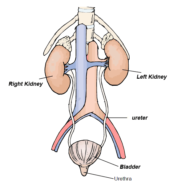
Urinary tract infections (UTIs) are common and usually occur because of the entry of bacteria into the urinary tract at the urethra The two forms of lower urinary tract infection (UTI) are cystitis (infection of the bladder) and urethritis (infection of the urethra). Urinary tract infection (UTI) more common in females than in males. UTI is prevalent in girls. In adult males and in children, lower UTIs typically are associated with anatomic or physiologic abnormalities and therefore need close evaluation. Most UTIs respond eadily to treatment, but recurrence and resistant bacteria flare-up during therapy are possible. 
“Urinary tract infection (UTI) is a bacterial infection that affects any part of the urinary tract. The main etiologic agent is Escherichia coli. Although urine contains a variety of fluids, salts, and waste products, it does not usually have bacteria in it. When bacteria get into the bladder or kidney and multiply in the urine, they may cause a UTI. “Pathogenesis 
The most common organism implicated in is E. coli and Staphylococcus. The bladder wall is coated with various mannosylated proteins, such as Tamm-Horsfall proteins (THP), which interfere with the binding of bacteria to the uroepithelium. As binding is an important factor in establishing pathogenicity for these organisms, its disruption results in reduced capacity for invasion of the tissues.[clarification needed] Moreover, the unbound bacteria are more easily removed when voiding. The use of urinary catheters (or other physical trauma) may physically disturb this protective lining, thereby allowing bacteria to invade the exposed epithelium. During cystitis, uropathogenic Escherichia coli (UPEC) subvert innate defenses by invading superficial umbrella cells and rapidly increasing in numbers to form intracellular bacterial communities (IBCs). By working together, bacteria in biofilms build themselves into structures that are more firmly anchored in infected cells and are more resistant to immune system assaults and antibiotic treatments This is often the cause of chronic urinary tract infections. 
Source: http://en.wikipedia.org/wiki/Urinary_tract_infection 

Urinary reflux is one reason that bacteria spread in the urinary tract. Vesicourethral reflux occurs when pressure increases in the bladder from coughing or sneezing and pushes urine into the urethra. When pressure returns to normal, the urine moves back into the bladder, taking with it bacteria from the urethra. In vesicoureteral reflux, urine flows backward from the bladder into one or both of the ureters, carrying bacteria from the bladder to the ureters and widening the infection. If they are left untreated, UTIs can lead to chronic infections, pyelonephritis, and even systemic sepsis and septic shock. If infection reaches the kidneys, permanent renal damage can occur, which leads to acute and chronic renal failure. 

Causes for Urinary tract infection (UTI) 
Most lower Urinary tract infection (UTI) result from ascending infection by a single gram-negative, enteric bacterium, such as Escherichia coli, Klebsiella, Proteus, Enterobacter, Pseudomonas, and Serratia. In a patient with neurogenic bladder, an indwelling urinary catheter, or a fistula between the intestine and bladder, a lower UTI may result from simultaneous infection with multiple pathogens. 
Studies suggest that infection results from a breakdown in local defense mechanisms in the bladder that allows bacteria to invade the bladder mucosa and multiply. These bacteria can’t be readily eliminated by normal urination. 
The pathogen’s resistance to the prescribed antimicrobial therapy usually causes bacterial flare-up during treatment. Even a small number of bacteria in a midstream urine specimen obtained during treatment casts doubt on the effectiveness of treatment. 
In almost all patients, recurrent lower Urinary tract infection (UTI) result from reinfection by the same organism or by some new pathogen. In the remaining patients, recurrence reflects persistent infection, usually from renal calculi, chronic bacterial prostatitis, or a structural anomaly that is a source of infection. The high incidence of lower UTI among females probably occurs because natural anatomic features that facilitate Urinary tract infection (UTI). 
Urinary tract infections (UTIs) are common and usually occur because of the entry of bacteria into the urinary tract at the urethra 

Nursing Assessment 
Patients History. The patient with a UTI has a variety of symptoms that range from mild to severe. The typical complaint is of one or more of the following: frequency, burning, urgency, nocturia, blood or pus in the urine, and suprapubic fullness. The patient may complain of urinary urgency and frequency, dysuria, bladder cramps or spasms, itching, a feeling of warmth during urination, nocturia. Other complaints include low back pain, malaise, nausea, vomiting, pain or tenderness over the bladder, chills, and flank pain. Inflammation of the bladder wall also causes hematuria and fever. Ask the patient about risk factors, including recent catheterization of the urinary tract, pregnancy or recent childbirth, neurological problems, volume depletion, frequent sexual activity, and presence of a sexually transmitted infection (STI). 
Physical Examination. Physical examination is often unremarkable in the patient with a UTI, although some patients have costovertebral angle tenderness in cases of pyelonephritis. On occasion, the patient has fever, chills, and signs of a systemic infection. Inspect the urine to determine its color, clarity, odor, and character. Surveillance for STIs is recommended as part of the examination. 

Diagnostic tests
Several tests are used to diagnose lower UTIs: Leukocyte esterase dip test Clean-catch urinalysis. Clean-catch collection is preferred to catheterization, which can reinfect the bladder with urethral bacteria. Sensitivity testing is used to determine the appropriate antimicrobial drug. Stained smear of urethral discharge can be used to rule out sexually transmitted disease. Voiding cystourethrography or excretory urography 

Nursing diagnosis
  • Acute pain 
  • Deficient knowledge (prevention) 
  • Disturbed sleep pattern 
  • Impaired urinary elimination 
  • Risk for infection 
  • Risk for injury Sexual dysfunction 


Nursing Key outcomes Nursing care plans for Urinary tract infections (UTIs) 
The patients will: Report increased comfort. Identify risk factors that exacerbate the disease process or condition and modify his lifestyle accordingly. Verbalize feeling well rested after undisturbed periods of sleep. Remain free from signs or symptoms of infection. Avoid or minimize complications. Reestablish sexual activity at the preillness level. Patient and family will demonstrate skill in managing elimination problem. 

Nursing interventions 
Administer antibiotics specific to the invading organism as ordered Watch for GI disturbances from antimicrobial therapy. If ordered, administer nitrofurantoin macrocrystals with milk or meals to prevent such distress. If the patient experiences perineal discomfort, sitz baths to the perineum may increase comfort. If sitz baths don’t relieve perineal discomfort, apply warm compresses sparingly to the perineum, but be careful not to burn the patient. Apply topical antiseptics on the urethral meatus as necessary. Collect urine specimens for culture and sensitivity testing carefully and promptly. Encourage patients to increase fluid intake to promote frequent urination 

Patient Teaching and Home Healthcare Guidelines
Teach the patient an understanding of the proposed therapy, including the medication name, dosage, route, and side effects, Emphasize the importance of completing the prescribed course of therapy or, with long-term prophylaxis, of strictly adhering to the ordered dosage. Explain that an uncontaminated midstream urine specimen is essential for accurate diagnosis. Teach the female patient to clean the perineum properly and to keep the labia separated during urination. To prevent recurrent lower UTIs, teach a female patient to carefully wipe the perineum from front to back and to thoroughly clean it with soap and water after bowel movements. Teach to the patients never to postpone urination and to empty her bladder completely. Tell the male patient that prompt treatment of predisposing conditions such as chronic prostatitis helps prevent recurrent UTIs. Urge the patient to drink about 2 qt (2 L) of fluid a day during treatment.