Wednesday, July 14, 2010

Bone Fractures X-ray
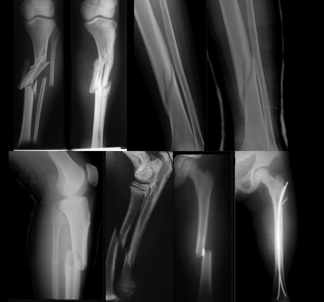
A fracture, or discontinuity of the bone, is the most common type of bone lesion. Normal bone can withstand considerable compression and shearing forces and, to a lesser extent, tension forces. A fracture occurs when more stress is placed on the bone than it is able to absorb.
A bone fracture is a medical condition in which there is a break in the continuity of the bone. A bone fracture can be the result of high force impactor stress, or trivial injury as a result of certain medical conditions that weaken the bones, such asosteoporosis, bone cancer, or osteogenesis imperfecta, where the fracture is then termedpathological fracture. (http://en.wikipedia.org/wiki/Bone_fracture)

Cause for Bone Fractures Bone Fractures Grouped according to cause, fractures can be divided into three major categories:

  1. Fractures caused by sudden injury The most common fractures result from major trauma, such as a fall on an outstretched arm, a skiing or motor vehicle accident, and child, spouse, or elder abuse (shown by multiple or repeated episodes of fractures). The force causing the fracture may be direct, such as a fall, or indirect, such as a massive muscle contraction or trauma transmitted along the bone. For example, the head of the radius or clavicle can be fractured by the indirect forces that result from falling on an outstretched hand. 
  2. Fatigue or stress fractures A fatigue fracture results from repeated wear on a bone. Pain associated with overuse injuries of the lower extremities, especially posterior medial tibial pain, is one of the most common symptoms that physically active persons, such as runners, experience Stress fractures in the tibia. 
  3. Pathologic fractures. A pathologic fracture is fracture that occurs when the normal integrity and strength of bone have been compromised by invasive disease or destructive processes or tumors. Fractures of this type may occur spontaneously with little or no stress. The underlying disease state can be local, as with infections, cysts, or tumors, or it can be generalized, as in osteoporosis, Paget’s disease, or disseminated tumors. 


Classification of Bone Fractures Fractures usually are classified according to: Location Type Direction or pattern of the fracture line.
Classification of Bone Fractures


Fragment position

  • Angulated, Bone fragments are at an angle to each other 
  • Avulsed, Bone fragments are pulled from normal position by muscle spasms, muscle contractions, or ligament resistance 
  • Comminuted, Bone breaks into many small pieces 
  • Displaced, Bone fragments separate and are deformed Impacted, A bone fragment is forced into another bone or bone fragment 
  • Nondisplaced, After the fracture, two sections of the bone maintain normal alignment 
  • Overriding, Bone fragments overlap, thereby shortening the total length of the bone
  • Segmental 


Fracture line

  • Linear Fracture line is parallel to the axis of the bone 
  • Longitudinal Fracture line extends longitudinally but not parallel to the axis of the bone 
  • Oblique Fracture line crosses the bone at a 45-degree angle to the axis of the bone 
  • Spiral Fracture line coils around the bone 
  • Transverse Fracture line forms a 90-degree angle to the axis of the bone 

A fracture of the long bone is described in relation to its position in the boneproximal, midshaft, and distal. Other descriptions are used when the fracture affects the head or neck of a bone, involves a joint, or is near a prominence such as a condyle or malleolus. The type of fracture is determined by its communication with the external environment, the degree of break in continuity of the bone, and the character of the fracture pieces.10
A fracture can be classified as open or closed. When the bone fragments have broken through the skin, the fracture is called an open or compound fracture. In a closed fracture, there is no communication with the outside skin.
The degree of a fracture is described in terms of a partial or complete break in the continuity of bone. A greenstick fracture, which is seen in children, is an example of a partial break in bone continuity and resembles that seen when a young sapling is broken. This kind of break occurs because children’s bones, especially until approximately 10 years of age, are more resilient than the bones of adults.
The character of the fracture pieces may also be used to describe a fracture. A comminuted fracture has more than two pieces. A compression fracture, as occurs in the vertebral body, involves two bones that are crushed or squeezed together. A fracture is called impacted when the fracture fragments are wedged together. This type usually occurs in the humerus, often is less serious, and usually is treated without surgery. Segmental fracture Bone fractures occur in two areas next to each other with an isolated section in the center
The direction of the trauma produces a certain configuration or pattern of fracture. Reduction is the restoration of a fractured bone to its normal anatomic position. The pattern of a fracture indicates the nature of the trauma and provides information about the easiest method for reduction. Linear fractures, Fracture line is parallel to the axis of the bone Transverse fractures are caused by simple angulatory forces. A spiral fracture results from a twisting motion, or torque. A transverse fracture is not likely to become displaced

Treatment for Bone Fractures
The primary goals of treatment are to return the injured limb to maximal function, to prevent complications, and to obtain the best possible cosmetic results. Emergency treatment consists of splinting the limb above and below the suspected fracture where it lies, applying a cold pack, and elevating the limb, all of which reduce edema and pain. A severe fracture that causes blood loss calls for direct pressure to control bleeding. The patient with a severe fracture may also need fluid replacement (including blood products) to prevent or treat hypovolemic shock.

Treatment Options for Bone Fractures
Treatment Options to set a Bone Fractures depends on the location and severity of the injury. To heal a bone fractures properly, the fractured bone must be realigned. The most common realignment procedures are: Immobilization using a cast or splint Setting of bone through surgery. Advantages of surgery include: early mobility of injured bone and some use of the injured bone within weeks rather than months. After the bone is realigned properly, medication and rehabilitation will help the recovery process. Medication is used to lessen the pain. Rehabilitation prevents stiffness. Rehabilitation involves light movement of the tissues surrounding the injury. It helps increase blood flow which will aid the healing process.

Nonoperative Management
Until comparatively recently, nonoperative treatment was the only method of treating fractures and severe soft tissue injuries, but the introduction of anesthesia, antibiotics, improved surgical implants, and better operative techniques has changed the treatment of many fractures. The process of change continues, and probably fewer fractures will be managed nonoperatively as the functional benefits of operative treatment become more apparent to both surgeons and patients.

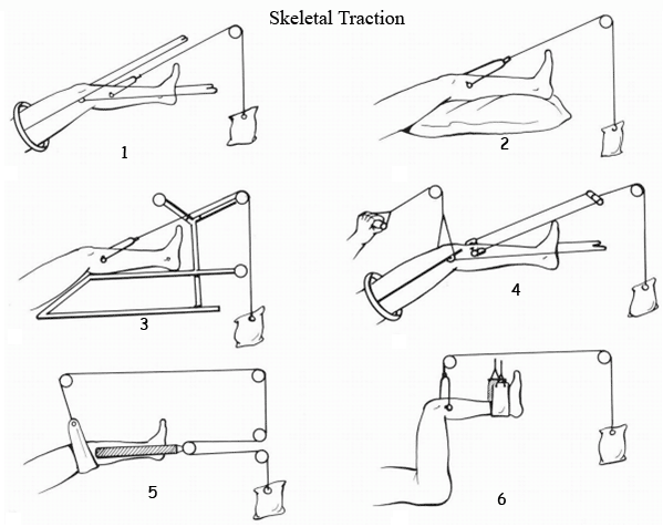
TRACTION: Skeletal Traction, Spinal Traction,
CASTS Braces
Slings, Bandages, and Support Strapping skeletal traction skeletal traction
skeletal traction


Operative Management
When closed reduction is impossible, open reduction during surgery use to reduces and immobilizes the fracture by means of rods, plates, or screws

  • Plating 
  • Intramedullary Nailing 
  • Kirschner wires 
  • External Fixation 
  • Arthroplasty 
  • Amputations 


Complications for Bone Fractures
Possible complications of fractures include arterial damage, nonunion, fat embolism, infection, shock, avascular necrosis, and peripheral nerve damage. Acute Compartment Syndrome Nonunions and Bone Defects Nursing diagnosis for bone fractures

Nursing diagnosis for bone fractures determine by data that we found in nursing assessment: Nursing Assessment nursing care plans for bone fractures
Assessment on patient’s history usually reveals what caused the fracture, major trauma, such as a fall on an outstretched arm, a skiing or motor vehicle accident, or elder abuse. The patient typically reports pain that increases with movement and an inability to move the part of the arm or leg distal to the injury. The severity of the pain depends on the fracture type. The patient may also complain of a tingling sensation distal to the injury, possibly indicating nerve and vessel damage.
Inspection may disclose soft-tissue edema, an obvious deformity or shortening of the injured limb, and discoloration over the fracture site. Open fractures produce an obvious skin wound and bleeding. Gentle palpation usually reveals warmth, crepitus and, possibly, dislocation. Numbness distal to the injury and cool skin at the end of the extremity may indicate nerve and vessel damage. Auscultation may reveal loss of pulses distal to the injury, an indication of possible arterial compromise or nerve damage.
Palpation pulses in distal of the fracture to detect injury to blood vessels, which is a surgical emergency

Diagnostic tests for Bone Fractures
Anteroposterior and lateral X-rays of the suspected fracture, as well as X-rays of the joints above and below it, confirm the diagnosis. Angiography can reveal concurrent vascular injury. MRI or CT Scan of spine if suspect a bone tumor or compression of spinal cord Bone densitometry can predict an increased risk of osteoporosis usually in pathologic fractures Blood tests

Nursing Care Plans for Bone Fractures. Common nursing diagnosis for bone fractures:

  • Acute pain, 
  • Anxiety, 
  • Bathing or hygiene self-care deficit, 
  • Fear, 
  • Impaired physical mobility, 
  • Ineffective coping, 
  • Ineffective role performance, 
  • Ineffective tissue perfusion: Peripheral, 
  • Risk for deficient fluid volume, 
  • Risk for disuse syndrome, 
  • Risk for infection, 
  • Risk for injury, 
  • Risk for [additional] Trauma. 


Nursing Goals Nursing Care Plans for Bone Fractures
Pain controlled. Prevented or minimized Complications Fracture stabilized. Condition, prognosis, and therapeutic regimen understood. Plan in place to meet needs after discharge. Nursing Care Plans for Bone Fractures with nursing diagnosis Acute pain

Sample Nursing Care Plans For bone Fracture with nursing diagnosis Pain Acute
NURSING
DIAGNOSIS
INTERVENTIONS
EVALUATION
Acute Pain related to Muscle spasms Movement of bone fragments, edema, and injury to the soft tissue
Traction, immobility device
Stress, anxiety
·       Perform a comprehensive assessment of pain including location, characteristics, onset/duration, frequency, quality, severity

·       Maintain immobilization of affected part


·       Elevate and support injured extremity

·       Perform and supervise passive or active ROM exercises

·       Suggest diversional activities appropriate for client’s age, physical abilities, and personal preferences


·       Administer medications, as indicated.
·       Verbalize relief of pain.

·       Follow prescribed pharmacologic regimen

·       Display relaxed manner, able to participate in activities, and sleep and rest appropriately

·       Demonstrate use of relaxation skills and diversional activities, as indicated for individual situation

Patient Teaching Discharge and Home Healthcare Guidelines for Fractures Patient 
Teaching Discharge and Home Health care Guidelines for fractures patient. To prevent complications of prolonged immobility, encourage the patient to participate in physical and occupational therapy as prescribed. Verify that the patient has demonstrated safe use of assistive devices such as wheelchairs, crutches, walkers, and transfers. Teach the patient the purpose, dosage, schedule, precautions, and potential side effects, interactions, and adverse reactions of all prescribed medications. Review with the patient all follow-up appointments that are arranged. If home care is necessary, verify that appropriate arrangements have been completed. 

  • Help the patient set realistic goals for recovery. 
  • Show the patient how to use his crutches properly. 
  • Tell the patient with a cast to report immediately signs of impaired circulation (skin coldness, numbness, tingling, or discoloration). 
  • Warn the patient against getting the cast wet, and instruct him not to insert foreign objects under the cast. Teach the patient to exercise joints above and below the cast as ordered. 
  • Tell the patient not to walk on a leg cast or foot cast without the physician’s permission. 
  • Emphasize the importance of returning for follow-up care.

Saturday, July 10, 2010

Asthma is a growing health problem, the number of children with asthma has increased markedly, unfortunately, and approximately 75% of children with asthma continue to have chronic problems in adulthood. Asthma is a reversible lung disease that may resolve spontaneously or with treatment, asthma is characterized by obstruction or narrowing of the airways, which are typically inflamed and hyperresponsive to various stimuli. Signs of asthma range from mild wheezing and Dyspnea to life-threatening respiratory failure. Symptoms of bronchial airway obstruction may persist between acute episodes. 
Hyper-reactivity leads to airway obstruction due to acute onset of muscle spasm in the smooth muscle of the tracheobronchial tree, thereby leading to a narrowed lumen. In addition to muscle spasm, there is swelling of the mucosa, which leads to edema. Lastly, the mucous glands increase in number, hypertrophy, and secrete thick mucus. 
In asthma, the total lung capacity (TLC), functional residual capacity (FRC), and residual volume (RV) increase, but the hallmark of airway obstruction is a reduction in ratio of the forced expiratory volume in 1 second (FEV1) and the FEV1 to the forced vital capacity (FVC). 
Although asthma can result from infections (especially viral) and inhaled irritants, it often is the result of an allergic response. An allergen (antigen) is introduced to the body, and sensitizing antibodies such as immunoglobulin E (IgE) are formed. IgE antibodies bind to tissue mast cells and basophils in the mucosa of the bronchioles, lung tissue, and nasopharynx. An antigen-antibody reaction releases primary mediator substances such as histamine and slow-reacting substance of anaphylaxis (SRS-A) and others. These mediators cause contraction of the smooth muscle and tissue edema. In addition, goblet cells secrete a thick mucus into the airways that causes obstruction. 

Extrinsic and intrinsic asthma 
For many asthmatics, intrinsic and extrinsic asthma coexist. Intrinsic asthma results from all other causes except allergies, such as infections (especially viral), inhaled irritants, and other causes or etiologies. The parasympathetic nervous system becomes stimulated, which increases bronchomotor tone, resulting in bronchoconstriction. 
Asthma that results from sensitivity to specific external allergens is referred to as extrinsic (atopic). In those cases where the allergen isn’t obvious, asthma is referred to as intrinsic (nonatopic). Allergens that cause extrinsic asthma include pollen, animal dander, house dust or mold, kapok or feather pillows, food additives containing sulfites, and any other sensitizing substance. 
Extrinsic asthma usually begins in childhood and is accompanied by other manifestations of atopy (type I, immunoglobulin [Ig] E–mediated allergy), such as eczema and allergic rhinitis. 
With intrinsic asthma, no extrinsic allergen can be identified. Most cases are preceded by a severe respiratory tract infection. Irritants, emotional stress, fatigue, exposure to noxious fumes, and endocrine, temperature, and humidity changes may aggravate intrinsic asthma attacks. 

Asthma Causes 
Asthma also called chronic reactive airway disease, chronic inflammatory disorder episodic exacerbations of reversible inflammation and hyperreactivity and variable constriction of bronchial smooth muscle, hypersecretion of mucus, and edema. Asthma that results from sensitivity to specific external allergens is known as extrinsic. In cases in which the allergen isn’t obvious, asthma is referred to as intrinsic. 
Extrinsic asthma Allergens include pollen, animal dander, house dust or mold, kapok or feather pillows, food additives containing sulfites, Genetic and environmental: household substances (such as dust mites, pets, cockroaches, mold), pollen, foods, latex, emotional upheaval, air pollution, cold weather, exercise, chemicals, medications, viral infections and any other sensitizing substance. 
Extrinsic asthma usually begins in childhood and is accompanied by other manifestations such as eczema and allergic rhinitis. In patients with intrinsic (nonatopic) asthma, no extrinsic allergen can be identified. Most cases are preceded by a severe respiratory tract infection. Irritants, emotional stress, fatigue, and exposure to noxious fumes, as well as endocrine changes and changes in temperature and humidity, may aggravate intrinsic asthma attacks. In many patients with asthma, intrinsic and extrinsic asthma coexist. Exercise may also provoke an asthma attack. In patients with exercise-induced asthma, bronchospasm may follow heat and moisture loss in the upper airways. 

Treatment for Asthma 
Treatment of acute asthma aims to decrease bronchoconstriction, reduce bronchial airway edema, and increase pulmonary ventilation. After an acute episode, treatment focuses on avoiding or removing precipitating factors, such as environmental allergens or irritants. 
Drug therapy is most effective when begun soon after the onset of symptoms. A patient who doesn’t respond to this treatment, whose airways remain obstructed, and who has increasing respiratory difficulty is at risk for status asthmaticus and may require mechanical ventilation. 

Nursing Assessment for patients with asthma 
An asthma attack may begin dramatically, with simultaneous onset of many severe symptoms, or insidiously, with gradually increasing respiratory distress. It typically includes progressively worsening shortness of breath, cough, wheezing, and chest tightness or some combination of these signs and symptoms. 
Patients history, obtain history of allergies thorough description of the response to allergens or other irritants. The patient may describe a sudden onset of symptoms after exposure, with a sense of suffocation. Symptoms include dyspnea, wheezing, and a cough and also chest tightness, restlessness, anxiety, and a prolonged expiratory phase. Ask if the patient has experienced a recent viral infection. 
Physical examination. severe shortness of breath can hardly speak, patients use their accessory muscles for breathing. Some patients have an increased anteroposterior thoracic diameter. If the patient has marked, color changes such as pallor or cyanosis or becomes confused, restless, or lethargic, increased risk of respiratory failure. 
Percussion of the lungs usually produces hyper-resonance, and palpation may reveal vocal fremitus. 
Auscultation high-pitched inspiratory and expiratory wheezes, prolonged expiratory phase of respiration. A rapid heart rate, mild systolic hypertension, and a paradoxic pulse may also be present. 

Diagnostic test for asthma 
Pulmonary function tests Pulse oximetry. Arterial blood gas (ABG) analysis. Complete blood count. Chest X-rays. Peak Expiratory Flow Rates (PEFR) 

Nursing diagnosis for Asthma 
Common nursing diagnosis found in Nursing Care Plans for Asthma; 
  • Impaired gas exchange related to Altered oxygen supply obstruction of airways by secretions, bronchospasm, air-trapping Alveoli destruction Ineffective airway clearance related to obstruction from narrowed lumen and thick mucus imbalanced 
  • Nutrition: Less than Body Requirements related to Dyspnea, sputum production Medication side effects; anorexia, nausea or vomiting 
  • Fatigue 
  • Ineffective breathing pattern 
  • Anxiety 
  • Deficient knowledge (treatment regimen, self-care, and discharge needs) 
  • Fear 

Sample Nursing care plans for Asthma
NURSING DIAGNOSIS
INTERVENTIONS
RATIONALE
EVALUATION
ineffective Airway Clearance R/T Bronchospasm
Increased production of secretions, retained secretions, thick, viscous secretions
Decreased energy or fatigue
·      Evaluate respiratory rate/depth and breath sounds.


·      Assist client to maintain a comfortable position.


·      Keep environmental free from sources of allergen such as dust, smoke, and feather pillows to a minimum according to individual situation.

·      Encourage/instruct in deep-breathing and directed coughing exercises


·     Tachypnea is usually present to some degree and may be pronounced during respiratory stress. 
·     facilitates respiratory function using gravity; however, client in severe distress will seek the position that most eases breathing

·     Precipitators of allergic type of respiratory reactions that can trigger or exacerbate onset of acute episode.
  
·     To maximize cough effort, lung expansion and drainage, and reduce pain impairment.
·        Respiratory Status: Airway Patency
·        Maintain patent airway with breath sounds clear or clearing.
·        Demonstrate behaviors to improve or maintain clear airway.
impaired Gas Exchange R/T Altered oxygen supply, obstruction of airways by secretions, bronchospasm
·      monitor skin and mucous membrane color.

·      Monitor vital signs
·      Encourage adequate rest and limit activities to within client tolerance.

·      Monitor and graph serial ABGs and pulse oximetry.

·      Administer medications as indicated
·     Duskiness and central cyanosis indicate advanced hypoxemia


·     Increased PaCO2 signals impending respiratory failure for asthmatics.
·        Demonstrate improved ventilation
·        Demonstrate adequate oxygenation of tissues by ABGs within client’s normal limits
·        absence of symptoms of respiratory distress

Patient Teaching Discharge and Home Healthcare Guidelines for Asthma
To prevent asthma attacks, teach patients the triggers that can precipitate an attack. Teach the patient and family the correct use of medications, including the dosage, route, action, and side Effects. In rare instances, asthma can lead to respiratory failure (status asthmaticus) if patients are not treated immediately or are unresponsive to treatment. Explain that any Dyspnea unrelieved by medications, and accompanied by wheezing and accessory muscle use, needs prompt attention from a healthcare provider. 
  • Teach the patient and his family to avoid known allergens and irritants. 
  • Teach the patient about his medications, drug interactions, including proper dosages, administration instructions, and adverse effects. 
  • Teach the patient how to use a metered dose inhaler. 
  • Explain how to use a peak flow meter to measure the degree of airway obstruction, If the patient has moderate to severe asthma. Tell him to keep a record and Explain the importance of calling the physician at once if the peak flow drops suddenly 
  • If the patient develops a fever above 100° F (37.8° C), chest pain, shortness of breath without coughing or exercising, or uncontrollable coughing. Tell the patient to notify the physician 
  • Teach the patient and his family an uncontrollable asthma attack requires immediate attention. 
  • Teach the patient diaphragmatic and effective coughing techniques. 
  • Urge him to Increase fluid intake to help loosen secretions and maintain hydration. 
  • Teach the patient and his family important of Regular medical follow-up care, when to notify healthcare professional of changes in condition, and periodic spirometry testing, chest x-rays, and sputum cultures.

Thursday, July 1, 2010

Anemia
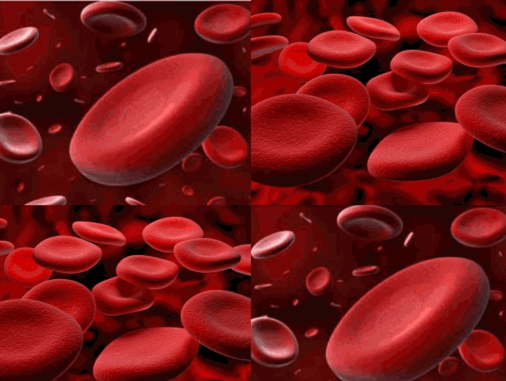
Iron deficiency anemia, anemia of chronic disease, pernicious anemia, Aplastic anemia, hemolytic anemia. The anemias are a group of blood disorders characterized by too little hemoglobin in the blood. Hemoglobin is a substance contained in red blood cells that carries oxygen from the lungs to other body tissues. Anemia is often a sign or symptom of an underlying disease rather than a disease in its own right. There are three tests commonly used to detect anemia: the number of red blood cells can be counted; the amount of hemoglobin in the red blood cells can be measured; or the proportion of blood cells to serum (the liquid part of blood, called the hematocrit) can be assessed. 
Adult anemia is usually defined as a Hemoglobin Hgb level lower than 11 g/dL, with severe anemia defined as Hemoglobin Hgb lower than 8 g/dL. Anemia Associated with many physiological complications, including dyspnea, fatigue, dizziness, decreased cognition, impaired sleep, sexual dysfunction, and significant debilitation 
Anemia can develop in three ways: loss of blood through injury, diseases of the digestive tract, or heavy menstrual flow in women; rapid destruction of red blood cells (e.g. sickle cell anemia); or inadequate production of healthy red blood cells (e.g. thalassemia). The underlying causes of anemias range from poor nutrition (iron-deficiency anemia) and digestive disorders (Crohn disease, celiac disease) to colorectal cancer, parasitic diseases (e.g. hookworm), and genetic disorders (sickle cell anemia, thalassemia). 

Pathophysiology: decreased number of circulating red blood cells (RBCs), reduction in the amount of hemoglobin (Hgb) in the RBCs, or a combination of both, resulting in diminished oxygen-carrying capacity of the blood 

  • Iron deficiency anemia inadequate iron stores, which results in insufficient Hemoglobin Hgb, causing cells to appear abnormal, unusually small (microcytic), and pale (hypochromic) 
  • Anemia of chronic disease; accompanies chronic inflammatory, infectious, or neoplastic disorders 
  • Pernicious anemia; lack of intrinsic factor in the stomach results in inability to absorb vitamin B12 causing abnormal RBC formation 
  • Aplastic anemia: failure of bone marrow to produce cells, including RBCs and white blood cells (WBCs) and platelets 
  • Hemolytic anemia: premature destruction of RBCs 


Causes and Complications of Anemia 
Causes and Etiology for Anemia, Anemia is often a sign or symptom of an underlying disease rather than a disease in its own. Anemia goes undetected in many people, and symptoms can be minor or vague. The signs and symptoms can be related to the anemia itself, or the underlying cause. 
Iron deficiency anemia Causes by Lack of iron in the body due to a variety of causes, inadequate nutrition, such as not enough foods that contain iron or Malabsorption syndromes. 
Anemia of chronic disease Primarily due to slowed production of RBCs because of low reticulocyte production. Symptoms usually associated with the disease causing the anemia rather than the anemia itself. 
Pernicious anemia An autoimmune disorder Characterized by the production of auto antibodies that destruct gastric parietal cells and their secretory product leads to a lack of intrinsic factor, which is needed for vitamin B12 absorption include Crohn’s and Whipple’s diseases, gastrectomy or gastric bypass, and chemotherapeutic medications. 
Aplastic anemia Bone marrow failure; May be associated with conditions that affect erythropoietin production and secretion, such as certain cancers and cancer treatments, hepatic, or endocrine disorders. Exposure to chemicals, immune conditions, such as systemic lupus erythematosus, or rheumatoid arthritis. 
Hemolytic anemia Accelerated destruction of RBCs sickle cell anemia Causes include hereditary factors, such as sickle cell trait or disease, blood transfusion reactions, acute viral or infectious agents, certain drugs, and toxins, such as chemicals and venoms. 

Complications of Anemia: 
Anemia is often a sign or symptom of an underlying disease rather than a disease in its own Mild anemia does not have any significant long-term consequences. As the anemia becomes more severe, medical problems may arise: High-output heart failure increased risk for a heart attack The lack of iron associated with anemia can cause many complications, including hypoxemia, brittle or rigid fingernails, cold intolerance, and possible behavioral disturbances in children. 

Nursing Assessment
Patient’s history because Anemia symptoms usually develop insidiously Patient’s history may not help to establish disease onset. The patient may report signs and symptoms of anemia (progressive weakness and fatigue, shortness of breath, and headache) or signs of thrombocytopenia (easy bruising and bleeding, especially from the mucous membranes) 
Inspection patients with Anemia may reveal pallor if the patient is anemic, and ecchymosis, petechiae, or retinal bleeding if thrombocytopenia is present. You may note alterations in the level of consciousness and weakness if bleeding into the central nervous system has occurred. 
Auscultation may reveal bibasilar crackles, tachycardia, and a gallop murmur if severe anemia results in heart failure. Fever, oral and rectal ulcers and sore throat may indicate the presence of an infection but without characteristic inflammation due to leukopenia. 

Diagnostic test for Anemia 
Complete blood count (CBC): Hgb; hematocrit (Hct); RBC count, morphology, indices, and distribution width index; platelet count and size; and WBC count and differential. Bone marrow biopsies 

Nursing diagnosis for Anemia 
Common nursing diagnosis found in Nursing care plans for anemia:

  • Activity Intolerance related to Imbalance between oxygen supply or delivery and demand 
  • Impaired oral mucous membrane 
  • Imbalanced Nutrition: Less than Body Requirements related to Failure to ingest or inability to digest food or absorb nutrients necessary for formation of normal RBCs 
  • Constipation/Diarrhea related to Decreased dietary intake, changes in digestive processes Drug therapy side effects 
  • Risk for Infection Inadequate secondary defenses—decreased Hgb, leukopenia, or decreased granulocytes (suppressed inflammatory response) Inadequate primary defenses—broken skin, stasis of body fluids, invasive procedures, chronic disease, malnutrition 
  • Risk for deficient fluid volume 
  • Deficient Knowledge regarding condition, prognosis, treatment, self-care, prevention of crisis, and discharge needs related to Lack of exposure, recall Information misinterpretation Unfamiliarity with information resource 
  • Fatigue 
  • Fear 
  • Ineffective coping
  • Ineffective thermoregulation 


Anemia are a group of blood disorders characterized by too little hemoglobin in the blood. Hemoglobin is a substance contained in red blood cells that carries oxygen from the lungs to other body tissues. Anemia is often a sign or symptom of an underlying disease rather than a disease in its own right. There are three tests commonly used to detect anemia: the number of red blood cells can be counted; the amount of hemoglobin in the red blood cells can be measured; or the proportion of blood cells to serum (the liquid part of blood, called the hematocrit) can be assessed. 

Sample Nursing care plans for anemia

NURSING DIAGNOSIS
INTERVENTIONS
RATIONALE
EVALUATION
Activity Intolerance related to Imbalance between oxygen supply or delivery and demand
·      Assess patient ability to perform ADLs


·      Monitor vital sign (Blood Pressure, pulse, and respirations) during and after activity


·      Suggest client change position slowly; monitor for dizziness.


·      Provide or recommend assistance with activities and ambulation as necessary, allowing client to be an active participant as much as possible.

·      Identify and implement energy-saving techniques


·      Instruct client to stop activity if palpitations, chest pain, shortness of breath, weakness, or dizziness occur


Collaborative
·      Monitor laboratory studies, such as Hgb/Hct, RBC count, and arterial blood gases (ABGs).


·      Provide supplemental oxygen as indicated.

  
·      Administer the following, as indicated: Whole blood, packed RBCs (PRCs); blood products as indicated.

·      Monitor closely for transfusion reactions.


·      Prepare for surgical intervention, if indicated.


·      Influences choice of interventions and needed assistance.

·      Cardiopulmonary manifestations result from attempts by the heart and lungs to supply adequate amounts of oxygen to the tissues.

·      Postural hypotension or cerebral hypoxia may cause dizziness, fainting, and increased risk of injury.

·      Although help may be necessary, self-esteem is enhanced when client does some things for self.

   
·      Encourages client to do as much as possible, while conserving limited energy and preventing fatigue.

·      Cellular ischemia potentiates risk of infarction, and excessive cardiopulmonary strain and stress may lead to decompensation and failure


·      Identifies deficiencies in RBC components affecting oxygen transport, treatment needs, and response to therapy.
·      Maximizing oxygen transport to tissues improves ability to function
·      Increases number of oxygen-carrying cells; corrects deficiencies to reduce risk of hemorrhage in acutely compromised individuals.


·      Surgery is useful to control bleeding in clients who are anemic because of bleeding, such as in ulcers and uterine bleeding; or to remove spleen as treatment of autoimmune hemolytic anemia. Bone marrow and stem cell transplantation may be done in presence of bone marrow failure aplastic anemia.

Report an increase in activity tolerance, including ADLs.

Demonstrate a decrease in physiological signs of intolerance pulse, respirations, and BP remain within client’s normal range.

Display laboratory values (Hgb/Hct) within acceptable range.

Patient Teaching and Home Healthcare Guidelines for Anemia
  • Teach the patient to avoid contact with potential sources of infection which can harbor organisms. 
  • Reassure and support the patient and his family by explaining the disease and its treatment, particularly if the patient has recurring acute episodes. 
  • Explain the purpose of all prescribed drugs, and discuss possible adverse reactions, including those he should report promptly. 
  • Tell the patient who does not require hospitalization that he can continue his normal lifestyle with appropriate restrictions. 
  • To prevent folic acid deficiency anemia, emphasize the importance of a well balanced diet high in folic acid. Teach the patient to meet daily folic acid requirements by including a food from each food group in every meal, Advise the patient not to stop taking the supplements when he begins to feel better. 
  • To help prevent exacerbation of sickle cell anemia, advise the patient to avoid tight clothing that restricts circulation. 
  • Emphasize the need for prompt treatment of infection.
  • Explain the need to increase fluid intake to prevent dehydration that results from impaired ability to properly concentrate urine. Tell parents to encourage a child with sickle cell anemia to drink more fluids. 
  • To encourage normal mental and social development, warn parents against being overprotective. Although the child must avoid strenuous exercise, he can enjoy most everyday activities. 
  • Refer parents of children with sickle cell anemia for genetic counseling to answer their questions about the risk to future offspring. Recommend screening of other family members to determine if they are heterozygote carriers. 
  • In sickle cell anemia Inform the patient and his parents that if he must be hospitalized for a vaso-occlusive crisis, I.V. fluids and a parenteral analgesic may be administered. He may also receive oxygen therapy and blood transfusions. 
  • Women with sickle cell anemia Warn them that they are poor obstetric risks. 
  • Emphasize the need for preventing trauma, abrasions, and breakdown of the skin. 
  • Be sure the patient understands the need to maintain a good nutritional intake to enhance the immune system and resistance to infections. 
  • Teach the patient the potential for bleeding and hemorrhage, and instruction to prevent bleeding. 
  • Discuss the need for regular dental examinations. 
  • Explain the importance of maintaining regular bowel movements to prevent straining.