Saturday, July 10, 2010

Asthma is a growing health problem, the number of children with asthma has increased markedly, unfortunately, and approximately 75% of children with asthma continue to have chronic problems in adulthood. Asthma is a reversible lung disease that may resolve spontaneously or with treatment, asthma is characterized by obstruction or narrowing of the airways, which are typically inflamed and hyperresponsive to various stimuli. Signs of asthma range from mild wheezing and Dyspnea to life-threatening respiratory failure. Symptoms of bronchial airway obstruction may persist between acute episodes. 
Hyper-reactivity leads to airway obstruction due to acute onset of muscle spasm in the smooth muscle of the tracheobronchial tree, thereby leading to a narrowed lumen. In addition to muscle spasm, there is swelling of the mucosa, which leads to edema. Lastly, the mucous glands increase in number, hypertrophy, and secrete thick mucus. 
In asthma, the total lung capacity (TLC), functional residual capacity (FRC), and residual volume (RV) increase, but the hallmark of airway obstruction is a reduction in ratio of the forced expiratory volume in 1 second (FEV1) and the FEV1 to the forced vital capacity (FVC). 
Although asthma can result from infections (especially viral) and inhaled irritants, it often is the result of an allergic response. An allergen (antigen) is introduced to the body, and sensitizing antibodies such as immunoglobulin E (IgE) are formed. IgE antibodies bind to tissue mast cells and basophils in the mucosa of the bronchioles, lung tissue, and nasopharynx. An antigen-antibody reaction releases primary mediator substances such as histamine and slow-reacting substance of anaphylaxis (SRS-A) and others. These mediators cause contraction of the smooth muscle and tissue edema. In addition, goblet cells secrete a thick mucus into the airways that causes obstruction. 

Extrinsic and intrinsic asthma 
For many asthmatics, intrinsic and extrinsic asthma coexist. Intrinsic asthma results from all other causes except allergies, such as infections (especially viral), inhaled irritants, and other causes or etiologies. The parasympathetic nervous system becomes stimulated, which increases bronchomotor tone, resulting in bronchoconstriction. 
Asthma that results from sensitivity to specific external allergens is referred to as extrinsic (atopic). In those cases where the allergen isn’t obvious, asthma is referred to as intrinsic (nonatopic). Allergens that cause extrinsic asthma include pollen, animal dander, house dust or mold, kapok or feather pillows, food additives containing sulfites, and any other sensitizing substance. 
Extrinsic asthma usually begins in childhood and is accompanied by other manifestations of atopy (type I, immunoglobulin [Ig] E–mediated allergy), such as eczema and allergic rhinitis. 
With intrinsic asthma, no extrinsic allergen can be identified. Most cases are preceded by a severe respiratory tract infection. Irritants, emotional stress, fatigue, exposure to noxious fumes, and endocrine, temperature, and humidity changes may aggravate intrinsic asthma attacks. 

Asthma Causes 
Asthma also called chronic reactive airway disease, chronic inflammatory disorder episodic exacerbations of reversible inflammation and hyperreactivity and variable constriction of bronchial smooth muscle, hypersecretion of mucus, and edema. Asthma that results from sensitivity to specific external allergens is known as extrinsic. In cases in which the allergen isn’t obvious, asthma is referred to as intrinsic. 
Extrinsic asthma Allergens include pollen, animal dander, house dust or mold, kapok or feather pillows, food additives containing sulfites, Genetic and environmental: household substances (such as dust mites, pets, cockroaches, mold), pollen, foods, latex, emotional upheaval, air pollution, cold weather, exercise, chemicals, medications, viral infections and any other sensitizing substance. 
Extrinsic asthma usually begins in childhood and is accompanied by other manifestations such as eczema and allergic rhinitis. In patients with intrinsic (nonatopic) asthma, no extrinsic allergen can be identified. Most cases are preceded by a severe respiratory tract infection. Irritants, emotional stress, fatigue, and exposure to noxious fumes, as well as endocrine changes and changes in temperature and humidity, may aggravate intrinsic asthma attacks. In many patients with asthma, intrinsic and extrinsic asthma coexist. Exercise may also provoke an asthma attack. In patients with exercise-induced asthma, bronchospasm may follow heat and moisture loss in the upper airways. 

Treatment for Asthma 
Treatment of acute asthma aims to decrease bronchoconstriction, reduce bronchial airway edema, and increase pulmonary ventilation. After an acute episode, treatment focuses on avoiding or removing precipitating factors, such as environmental allergens or irritants. 
Drug therapy is most effective when begun soon after the onset of symptoms. A patient who doesn’t respond to this treatment, whose airways remain obstructed, and who has increasing respiratory difficulty is at risk for status asthmaticus and may require mechanical ventilation. 

Nursing Assessment for patients with asthma 
An asthma attack may begin dramatically, with simultaneous onset of many severe symptoms, or insidiously, with gradually increasing respiratory distress. It typically includes progressively worsening shortness of breath, cough, wheezing, and chest tightness or some combination of these signs and symptoms. 
Patients history, obtain history of allergies thorough description of the response to allergens or other irritants. The patient may describe a sudden onset of symptoms after exposure, with a sense of suffocation. Symptoms include dyspnea, wheezing, and a cough and also chest tightness, restlessness, anxiety, and a prolonged expiratory phase. Ask if the patient has experienced a recent viral infection. 
Physical examination. severe shortness of breath can hardly speak, patients use their accessory muscles for breathing. Some patients have an increased anteroposterior thoracic diameter. If the patient has marked, color changes such as pallor or cyanosis or becomes confused, restless, or lethargic, increased risk of respiratory failure. 
Percussion of the lungs usually produces hyper-resonance, and palpation may reveal vocal fremitus. 
Auscultation high-pitched inspiratory and expiratory wheezes, prolonged expiratory phase of respiration. A rapid heart rate, mild systolic hypertension, and a paradoxic pulse may also be present. 

Diagnostic test for asthma 
Pulmonary function tests Pulse oximetry. Arterial blood gas (ABG) analysis. Complete blood count. Chest X-rays. Peak Expiratory Flow Rates (PEFR) 

Nursing diagnosis for Asthma 
Common nursing diagnosis found in Nursing Care Plans for Asthma; 
  • Impaired gas exchange related to Altered oxygen supply obstruction of airways by secretions, bronchospasm, air-trapping Alveoli destruction Ineffective airway clearance related to obstruction from narrowed lumen and thick mucus imbalanced 
  • Nutrition: Less than Body Requirements related to Dyspnea, sputum production Medication side effects; anorexia, nausea or vomiting 
  • Fatigue 
  • Ineffective breathing pattern 
  • Anxiety 
  • Deficient knowledge (treatment regimen, self-care, and discharge needs) 
  • Fear 

Sample Nursing care plans for Asthma
NURSING DIAGNOSIS
INTERVENTIONS
RATIONALE
EVALUATION
ineffective Airway Clearance R/T Bronchospasm
Increased production of secretions, retained secretions, thick, viscous secretions
Decreased energy or fatigue
·      Evaluate respiratory rate/depth and breath sounds.


·      Assist client to maintain a comfortable position.


·      Keep environmental free from sources of allergen such as dust, smoke, and feather pillows to a minimum according to individual situation.

·      Encourage/instruct in deep-breathing and directed coughing exercises


·     Tachypnea is usually present to some degree and may be pronounced during respiratory stress. 
·     facilitates respiratory function using gravity; however, client in severe distress will seek the position that most eases breathing

·     Precipitators of allergic type of respiratory reactions that can trigger or exacerbate onset of acute episode.
  
·     To maximize cough effort, lung expansion and drainage, and reduce pain impairment.
·        Respiratory Status: Airway Patency
·        Maintain patent airway with breath sounds clear or clearing.
·        Demonstrate behaviors to improve or maintain clear airway.
impaired Gas Exchange R/T Altered oxygen supply, obstruction of airways by secretions, bronchospasm
·      monitor skin and mucous membrane color.

·      Monitor vital signs
·      Encourage adequate rest and limit activities to within client tolerance.

·      Monitor and graph serial ABGs and pulse oximetry.

·      Administer medications as indicated
·     Duskiness and central cyanosis indicate advanced hypoxemia


·     Increased PaCO2 signals impending respiratory failure for asthmatics.
·        Demonstrate improved ventilation
·        Demonstrate adequate oxygenation of tissues by ABGs within client’s normal limits
·        absence of symptoms of respiratory distress

Patient Teaching Discharge and Home Healthcare Guidelines for Asthma
To prevent asthma attacks, teach patients the triggers that can precipitate an attack. Teach the patient and family the correct use of medications, including the dosage, route, action, and side Effects. In rare instances, asthma can lead to respiratory failure (status asthmaticus) if patients are not treated immediately or are unresponsive to treatment. Explain that any Dyspnea unrelieved by medications, and accompanied by wheezing and accessory muscle use, needs prompt attention from a healthcare provider. 
  • Teach the patient and his family to avoid known allergens and irritants. 
  • Teach the patient about his medications, drug interactions, including proper dosages, administration instructions, and adverse effects. 
  • Teach the patient how to use a metered dose inhaler. 
  • Explain how to use a peak flow meter to measure the degree of airway obstruction, If the patient has moderate to severe asthma. Tell him to keep a record and Explain the importance of calling the physician at once if the peak flow drops suddenly 
  • If the patient develops a fever above 100° F (37.8° C), chest pain, shortness of breath without coughing or exercising, or uncontrollable coughing. Tell the patient to notify the physician 
  • Teach the patient and his family an uncontrollable asthma attack requires immediate attention. 
  • Teach the patient diaphragmatic and effective coughing techniques. 
  • Urge him to Increase fluid intake to help loosen secretions and maintain hydration. 
  • Teach the patient and his family important of Regular medical follow-up care, when to notify healthcare professional of changes in condition, and periodic spirometry testing, chest x-rays, and sputum cultures.
Thursday, July 1, 2010

Anemia
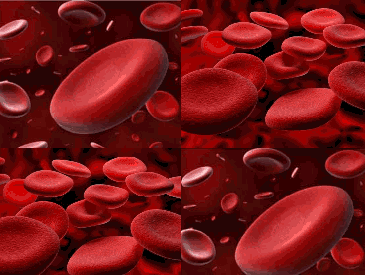
Iron deficiency anemia, anemia of chronic disease, pernicious anemia, Aplastic anemia, hemolytic anemia. The anemias are a group of blood disorders characterized by too little hemoglobin in the blood. Hemoglobin is a substance contained in red blood cells that carries oxygen from the lungs to other body tissues. Anemia is often a sign or symptom of an underlying disease rather than a disease in its own right. There are three tests commonly used to detect anemia: the number of red blood cells can be counted; the amount of hemoglobin in the red blood cells can be measured; or the proportion of blood cells to serum (the liquid part of blood, called the hematocrit) can be assessed. 
Adult anemia is usually defined as a Hemoglobin Hgb level lower than 11 g/dL, with severe anemia defined as Hemoglobin Hgb lower than 8 g/dL. Anemia Associated with many physiological complications, including dyspnea, fatigue, dizziness, decreased cognition, impaired sleep, sexual dysfunction, and significant debilitation 
Anemia can develop in three ways: loss of blood through injury, diseases of the digestive tract, or heavy menstrual flow in women; rapid destruction of red blood cells (e.g. sickle cell anemia); or inadequate production of healthy red blood cells (e.g. thalassemia). The underlying causes of anemias range from poor nutrition (iron-deficiency anemia) and digestive disorders (Crohn disease, celiac disease) to colorectal cancer, parasitic diseases (e.g. hookworm), and genetic disorders (sickle cell anemia, thalassemia). 

Pathophysiology: decreased number of circulating red blood cells (RBCs), reduction in the amount of hemoglobin (Hgb) in the RBCs, or a combination of both, resulting in diminished oxygen-carrying capacity of the blood 

  • Iron deficiency anemia inadequate iron stores, which results in insufficient Hemoglobin Hgb, causing cells to appear abnormal, unusually small (microcytic), and pale (hypochromic) 
  • Anemia of chronic disease; accompanies chronic inflammatory, infectious, or neoplastic disorders 
  • Pernicious anemia; lack of intrinsic factor in the stomach results in inability to absorb vitamin B12 causing abnormal RBC formation 
  • Aplastic anemia: failure of bone marrow to produce cells, including RBCs and white blood cells (WBCs) and platelets 
  • Hemolytic anemia: premature destruction of RBCs 


Causes and Complications of Anemia 
Causes and Etiology for Anemia, Anemia is often a sign or symptom of an underlying disease rather than a disease in its own. Anemia goes undetected in many people, and symptoms can be minor or vague. The signs and symptoms can be related to the anemia itself, or the underlying cause. 
Iron deficiency anemia Causes by Lack of iron in the body due to a variety of causes, inadequate nutrition, such as not enough foods that contain iron or Malabsorption syndromes. 
Anemia of chronic disease Primarily due to slowed production of RBCs because of low reticulocyte production. Symptoms usually associated with the disease causing the anemia rather than the anemia itself. 
Pernicious anemia An autoimmune disorder Characterized by the production of auto antibodies that destruct gastric parietal cells and their secretory product leads to a lack of intrinsic factor, which is needed for vitamin B12 absorption include Crohn’s and Whipple’s diseases, gastrectomy or gastric bypass, and chemotherapeutic medications. 
Aplastic anemia Bone marrow failure; May be associated with conditions that affect erythropoietin production and secretion, such as certain cancers and cancer treatments, hepatic, or endocrine disorders. Exposure to chemicals, immune conditions, such as systemic lupus erythematosus, or rheumatoid arthritis. 
Hemolytic anemia Accelerated destruction of RBCs sickle cell anemia Causes include hereditary factors, such as sickle cell trait or disease, blood transfusion reactions, acute viral or infectious agents, certain drugs, and toxins, such as chemicals and venoms. 

Complications of Anemia: 
Anemia is often a sign or symptom of an underlying disease rather than a disease in its own Mild anemia does not have any significant long-term consequences. As the anemia becomes more severe, medical problems may arise: High-output heart failure increased risk for a heart attack The lack of iron associated with anemia can cause many complications, including hypoxemia, brittle or rigid fingernails, cold intolerance, and possible behavioral disturbances in children. 

Nursing Assessment
Patient’s history because Anemia symptoms usually develop insidiously Patient’s history may not help to establish disease onset. The patient may report signs and symptoms of anemia (progressive weakness and fatigue, shortness of breath, and headache) or signs of thrombocytopenia (easy bruising and bleeding, especially from the mucous membranes) 
Inspection patients with Anemia may reveal pallor if the patient is anemic, and ecchymosis, petechiae, or retinal bleeding if thrombocytopenia is present. You may note alterations in the level of consciousness and weakness if bleeding into the central nervous system has occurred. 
Auscultation may reveal bibasilar crackles, tachycardia, and a gallop murmur if severe anemia results in heart failure. Fever, oral and rectal ulcers and sore throat may indicate the presence of an infection but without characteristic inflammation due to leukopenia. 

Diagnostic test for Anemia 
Complete blood count (CBC): Hgb; hematocrit (Hct); RBC count, morphology, indices, and distribution width index; platelet count and size; and WBC count and differential. Bone marrow biopsies 

Nursing diagnosis for Anemia 
Common nursing diagnosis found in Nursing care plans for anemia:

  • Activity Intolerance related to Imbalance between oxygen supply or delivery and demand 
  • Impaired oral mucous membrane 
  • Imbalanced Nutrition: Less than Body Requirements related to Failure to ingest or inability to digest food or absorb nutrients necessary for formation of normal RBCs 
  • Constipation/Diarrhea related to Decreased dietary intake, changes in digestive processes Drug therapy side effects 
  • Risk for Infection Inadequate secondary defenses—decreased Hgb, leukopenia, or decreased granulocytes (suppressed inflammatory response) Inadequate primary defenses—broken skin, stasis of body fluids, invasive procedures, chronic disease, malnutrition 
  • Risk for deficient fluid volume 
  • Deficient Knowledge regarding condition, prognosis, treatment, self-care, prevention of crisis, and discharge needs related to Lack of exposure, recall Information misinterpretation Unfamiliarity with information resource 
  • Fatigue 
  • Fear 
  • Ineffective coping
  • Ineffective thermoregulation 


Anemia are a group of blood disorders characterized by too little hemoglobin in the blood. Hemoglobin is a substance contained in red blood cells that carries oxygen from the lungs to other body tissues. Anemia is often a sign or symptom of an underlying disease rather than a disease in its own right. There are three tests commonly used to detect anemia: the number of red blood cells can be counted; the amount of hemoglobin in the red blood cells can be measured; or the proportion of blood cells to serum (the liquid part of blood, called the hematocrit) can be assessed. 

Sample Nursing care plans for anemia

NURSING DIAGNOSIS
INTERVENTIONS
RATIONALE
EVALUATION
Activity Intolerance related to Imbalance between oxygen supply or delivery and demand
·      Assess patient ability to perform ADLs


·      Monitor vital sign (Blood Pressure, pulse, and respirations) during and after activity


·      Suggest client change position slowly; monitor for dizziness.


·      Provide or recommend assistance with activities and ambulation as necessary, allowing client to be an active participant as much as possible.

·      Identify and implement energy-saving techniques


·      Instruct client to stop activity if palpitations, chest pain, shortness of breath, weakness, or dizziness occur


Collaborative
·      Monitor laboratory studies, such as Hgb/Hct, RBC count, and arterial blood gases (ABGs).


·      Provide supplemental oxygen as indicated.

  
·      Administer the following, as indicated: Whole blood, packed RBCs (PRCs); blood products as indicated.

·      Monitor closely for transfusion reactions.


·      Prepare for surgical intervention, if indicated.


·      Influences choice of interventions and needed assistance.

·      Cardiopulmonary manifestations result from attempts by the heart and lungs to supply adequate amounts of oxygen to the tissues.

·      Postural hypotension or cerebral hypoxia may cause dizziness, fainting, and increased risk of injury.

·      Although help may be necessary, self-esteem is enhanced when client does some things for self.

   
·      Encourages client to do as much as possible, while conserving limited energy and preventing fatigue.

·      Cellular ischemia potentiates risk of infarction, and excessive cardiopulmonary strain and stress may lead to decompensation and failure


·      Identifies deficiencies in RBC components affecting oxygen transport, treatment needs, and response to therapy.
·      Maximizing oxygen transport to tissues improves ability to function
·      Increases number of oxygen-carrying cells; corrects deficiencies to reduce risk of hemorrhage in acutely compromised individuals.


·      Surgery is useful to control bleeding in clients who are anemic because of bleeding, such as in ulcers and uterine bleeding; or to remove spleen as treatment of autoimmune hemolytic anemia. Bone marrow and stem cell transplantation may be done in presence of bone marrow failure aplastic anemia.

Report an increase in activity tolerance, including ADLs.

Demonstrate a decrease in physiological signs of intolerance pulse, respirations, and BP remain within client’s normal range.

Display laboratory values (Hgb/Hct) within acceptable range.

Patient Teaching and Home Healthcare Guidelines for Anemia
  • Teach the patient to avoid contact with potential sources of infection which can harbor organisms. 
  • Reassure and support the patient and his family by explaining the disease and its treatment, particularly if the patient has recurring acute episodes. 
  • Explain the purpose of all prescribed drugs, and discuss possible adverse reactions, including those he should report promptly. 
  • Tell the patient who does not require hospitalization that he can continue his normal lifestyle with appropriate restrictions. 
  • To prevent folic acid deficiency anemia, emphasize the importance of a well balanced diet high in folic acid. Teach the patient to meet daily folic acid requirements by including a food from each food group in every meal, Advise the patient not to stop taking the supplements when he begins to feel better. 
  • To help prevent exacerbation of sickle cell anemia, advise the patient to avoid tight clothing that restricts circulation. 
  • Emphasize the need for prompt treatment of infection.
  • Explain the need to increase fluid intake to prevent dehydration that results from impaired ability to properly concentrate urine. Tell parents to encourage a child with sickle cell anemia to drink more fluids. 
  • To encourage normal mental and social development, warn parents against being overprotective. Although the child must avoid strenuous exercise, he can enjoy most everyday activities. 
  • Refer parents of children with sickle cell anemia for genetic counseling to answer their questions about the risk to future offspring. Recommend screening of other family members to determine if they are heterozygote carriers. 
  • In sickle cell anemia Inform the patient and his parents that if he must be hospitalized for a vaso-occlusive crisis, I.V. fluids and a parenteral analgesic may be administered. He may also receive oxygen therapy and blood transfusions. 
  • Women with sickle cell anemia Warn them that they are poor obstetric risks. 
  • Emphasize the need for preventing trauma, abrasions, and breakdown of the skin. 
  • Be sure the patient understands the need to maintain a good nutritional intake to enhance the immune system and resistance to infections. 
  • Teach the patient the potential for bleeding and hemorrhage, and instruction to prevent bleeding. 
  • Discuss the need for regular dental examinations. 
  • Explain the importance of maintaining regular bowel movements to prevent straining.

Friday, June 18, 2010

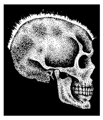
skull abnormality in a patient with thalassemia major
Thalassemia is a group of hereditary hemolytic anemia, which Marked by a deficit in the production of hemoglobin. Where the damage occurred in the red blood cells in the blood vessel so that the age erythrocyte be short. It is characterized by defective synthesis in one or more of the polypeptide chains necessary for hemoglobin production. Because Thalassemia affects hemoglobin production, it also impairs red blood cell (RBC) synthesis. 
There are two main types of Thalassemia: ”alpha” and “beta.” The names refer to the part of the hemoglobin molecule that is missing or mutated. 
Two pairs of polypeptide chains”alpha and beta chains” make up hemoglobin. In Thalassemia, diminished synthesis can affect either pair. Structurally, the chains are normal, but the genetic defect decreases their number. In alpha Thalassemia, alpha chain synthesis slows; in beta-Thalassemia, beta chain synthesis slows. Some patients with beta-Thalassemia have no normal hemoglobin only hemoglobin S and the minor hemoglobin’s. 

Causes for Thalassemia 
Thalassemia major Causes by homozygous inheritance of the partially dominant autosomal gene Thalassemia intermedia Causes by homozygous inheritance of the partially dominant autosomal gene. Thalassemia minor is caused by heterozygous inheritance of the same gene. In All type of Thalassemia deficiency of beta polypeptide chain production impairs hemoglobin synthesis and results in continual production of fetal hemoglobin, even after the neonatal period. 

Complications for Thalassemia 
Pathologic fractures Cardiac arrhythmias Heart failure Other complications that result from iron deposits in the heart and other tissues caused by repeated blood transfusions. 

Treatment for Thalassemia 
Patients with Thalassemia minor usually do not require any specific treatment. Treatment for patients with Thalassemia major includes chronic blood transfusion therapy, iron chelation, splenectomy, and allogeneic hematopoietic transplantation. References: http://en.wikipedia.org/wiki/Thalassemia 

Nursing Assessment
Thalassemia major: severe anemia, splenomegaly or hepatomegaly with abdominal enlargement, frequent infections, bleeding tendencies e.g. epistaxis, and anorexia, small bodies and large heads, mentally disabled, mongoloid. Thalassemia intermediate: anemia, jaundice, and splenomegaly, hemosiderosis caused by increased intestinal absorption of iron. Thalassemia minor: mild anemia usually with no signs or symptoms. 

Diagnostic Test for Thalassemia 
RBC count and hemoglobin level X-rays of the skull and long bones 

Nursing Diagnosis
Activity intolerance Deficient knowledge (treatment regimen) Delayed growth and development Disturbed body image Ineffective tissue perfusion: Cardiopulmonary Interrupted family processes Risk for infection 

Nursing care Plans for Thalassemia 
Treatment of thalassemia major is essentially supportive. For example, infections require prompt treatment with the appropriate antibiotic. Transfusions of packed RBCs raise the hemoglobin level but must be used judiciously to minimize iron overload. Thalassemia intermedia and thalassemia minor generally don’t require treatment. Iron supplements are contraindicated in all forms of thalassemia. Treatment of children is more difficult. Regular blood transfusions may minimize physical and mental retardation, but transfusions increase the risk of deadly hemosiderosis and iron overload. Continuous subcutaneous infusion of an iron-chelating agent may help produce a negative overall iron balance. If rapid splenic sequestration of transfused RBCs necessitates more transfusions, a splenectomy may be performed. 

Nursing outcomes 
Patient and parents will: Verbalize the importance of balancing activity, as tolerated, with frequent rest periods. Demonstrate age-appropriate skills and behaviors to the extent possible. Express positive feelings about himself. Develop no cardiac arrhythmias. Voice feelings and concerns related to the patient’s illness. Remain free from signs and symptoms of infection Nursing interventions nursing care plans for Thalassemia Watch for adverse reactions during and after RBC transfusions. Collaborative an antibiotic, and observe the patient for adverse reactions. Provide an adequate diet, Encourage the patient to drink plenty of fluids. Provide emotional support Help the patient and his family cope for chronic nature of Explain the need for lifelong transfusions. 

Patient teaching
Explain how to prevent infection e.g. nutrition, wound care Tell about signs of hepatitis and iron overload, which are always possible with frequent transfusions. Explain why child must avoid strenuous athletic activity to avoid pathologic fractures.

Friday, June 11, 2010

Acute Pain
NANDA Nursing Diagnosis for Acute Pain Related factors R/T trauma, injuring agents (biological, chemical, physical, psychological) 
Suggestion on using NANDA Nursing Diagnosis Acute Pain: Distinguish between acute pain Nursing diagnosis with the chronicles pain Nursing diagnosis. One of the two sets is that diagnosis of a painful time. ONSET acute pain is less than 6 months, while the painful chronicles ONSET it is more than 6 months. If you only have two diagnoses to indicate, pain is acute pain and chronic pain. Thus, there is no automatic diagnosis Crashes feel comfortable or feel comfortable painful chronic pain. 
Definition Nursing Diagnosis for Acute Pain: Unpleasant sensory and emotional experience arising from actual or potential tissue damage or described in terms of such damage (International Association for the Study of Pain); sudden or slow onset of any intensity from mild to severe with an anticipated or predictable end and a duration of less than 6 months 

Nursing Outcomes Client Will: Report pain is relieved / controlled. Follow prescribed pharmacological regimen. Verbalize methods that provide relief. Demonstrate use of relaxation skills and diversion activities as indicated for individual situation. 

Nursing Priority Nursing Diagnosis for Acute Pain To assess etiology/precipitating contributory factors: evaluate client’s response to pain: assist client to explore methods for alleviation/control of pain 

Sample Clinical Applications using Nursing Diagnosis for Acute Pain: Traumatic injuries, surgical procedures, infections, cancer, burns, skin lesions, gangrene, thrombophlebitis/pulmonary embolus, neuralgia

Thursday, June 10, 2010

Malignant Brain Tumors
Primary brain tumors comprise a diverse group of neoplasm’s that are often malignant and refractory to treatment, malignant brain tumors. More common in males than in females, malignant brain tumors (gliomas, meningiomas, and schwannomas) they cause central nervous system (CNS) changes by invading and destroying tissues and by secondary effects mainly compression of the brain, cranial nerves, and cerebral vessels; cerebral edema; and increased intracranial pressure ICP. Tumors can occur at any age. In adults, incidence is highest between ages 40 and 60, and the most common tumor types are gliomas and meningiomas. They usually occur above the covering of the cerebellum (supratentorial tumors). Most tumors in children occur before age one or between ages 2 and 12. The most common are astrocytomas, medulloblastomas, ependymomas, and brain stem gliomas. Brain tumors are one of the most common causes of cancer death in children. 

Primary brain tumors classified by their siteof origin 
  • CNS tumors that originate in glial tissue Gliomas, occurring in the glial, or supportive tissues around the brain: Astrocytomas Brain-stem gliomas Ependymomas Oligodendrogliomas 
  • CNS tumors that do not originate in glial tissue: Medulloblastoma Craniopharyngiomas Germinomas Pineal-region tumors 


Causes of Brain Tumor 
Brain tumors may be attributed to a familial link or to exposure to ionized radiation (a known environmental risk). Brain tumors may result from metastasis in 20% to 40% of patients with other cancers. In some cases, the cause remains unknown. 

Risk Factors of Primary Brain Tumors 
Hereditary syndromes (proven): tuberous sclerosis, neurofibromatosis types 1 and 2, nevoid basal cell carcinoma syndrome, Turcot’s syndrome, and Li-Fraumeni syndrome Family History of brain tumors History of prior cancer Exposure to infectious agents Allergies (possible reduced risk) Head trauma Drugs and medications Dietary history: N-nitroso compounds, oxidants, antioxidants Tobacco usage Alcohol consumption Ionizing radiation exposure Cellular telephones, Power frequency electromagnetic field exposure 

Complications 
In malignant brain tumors, life-threatening complications from increasing ICP include coma, respiratory or cardiac arrest, and brain herniation

Monday, June 7, 2010

Cervical cancer is the third most common cancer of the female reproductive system. Cancer of the cervix is one type of primary uterine cancer (the other being uterine-endometrial cancer) and is predominately epidermoid. Invasive cervical cancer is the third most common female pelvic cancer. The death rate from cervical cancer has steadily declined over the past 50 years owing to the increased use of the Papanicolaou exam, which detects cervical changes before cancer develops. Three types of cervical cancer are: Dysplasia, Carcinoma in situ (CIS) and Invasive carcinoma 
Preinvasive cancer ranges from minimal cervical dysplasia, in which the lower third of the epithelium contains abnormal cells, to carcinoma in situ, in which the full thickness of epithelium contains abnormally proliferating cells (also known as cervical intraepithelial neoplasia). Preinvasive cancer is curable in 75% to 90% of patients with early detection and proper treatment. If untreated, it may progress to invasive cervical cancer, depending on the form. 
CIS is carcinoma confined to the epithelium. The full thickness of the epithelium contains abnormally proliferating cells. Both dysplasia and CIS are considered preinvasive cancers and, with early detection, have a 5-year survival rate of 73% to 92%. 
In invasive disease, cancer cells penetrate the basement membrane and can spread directly to contiguous pelvic structures or disseminate to distant sites by way of lymphatic routes. In 95% of cases, the histologic type is squamous cell carcinoma, which varies from well-differentiated cells to highly anaplastic spindle cells. Only 5% of cases are adenocarcinomas. Invasive cancer typically occurs between ages 30 and 50; it rarely occurs younger than age 20. 

Cervical cancer stage (source: http://en.wikipedia.org) 
Cervical cancer is staged by the International Federation of Gynecology and Obstetrics (FIGO) staging system, which is based on clinical examination, rather than surgical findings. It allows only the following diagnostic tests to be used in determining the stage: palpation, inspection, colposcopy, endocervical curettage, hysteroscopy, cystoscopy, proctoscopy, intravenous urography, and X-ray examination of the lungs and skeleton, and cervical conization. 
The TNM staging system for cervical cancer is analogous to the FIGO stage. 

Stage 0 – full-thickness involvement of the epithelium without invasion into the stroma (carcinoma in situ) 
Stage I – limited to the cervix 
IA – diagnosed only by microscopy; no visible lesions 
          IA1 – stromal invasion less than 3 mm in depth and 7 mm or less in horizontal spread 
          IA2 – stromal invasion between 3 and 5 mm with horizontal spread of 7 mm or less 
IB – visible lesion or a microscopic lesion with more than 5 mm of depth or horizontal spread of more than 7 mm 
          IB1 – visible lesion 4 cm or less in greatest dimension 
          IB2 – visible lesion more than 4 cm 
Stage II – invades beyond cervix
          IIA – without parametrial invasion, but involve upper 2/3 of vagina
          IIB – with parametrial invasion 
Stage III – extends to pelvic wall or lower third of the vagina
          IIIA – involves lower third of vagina
          IIIB – extends to pelvic wall and/or causes hydronephrosis or non-functioning kidney 
IVA – invades mucosa of bladder or rectum and/or extends beyond true pelvis 
IVB – distant metastasis 

Causes for Cervical cancer 
Worldwide studies suggest that Causes for Cervical cancer is sexually transmitted human papillomaviruses (HPVs). Certain strains of HPV (16, 18, and 31) are associated with an increased risk of cervical cancer. Several predisposing factors have been related to the development of cervical cancer: intercourse at a young age), multiple sexual partners, and herpesvirus 2 and other bacterial or viral venereal infections. Genetic considerations While most risk factors for cervical cancer are environmental, some studies have found that the daughters or sisters of cervical cancer patients are more likely to get the disease. 

Complications of Cervical cancer 
Disease progression can cause flank pain from sciatic nerve or pelvic wall invasion and hematuria and renal failure associated with bladder involvement.

  • Ureteric obstruction 
  • Intermenstrual PV bleed 
  • Vesicovaginal fistula 
  • Post-menopausal PV bleed 
  • Uterine enlargement 
  • Menorrhagia 

Nursing Assessment
Patient’s history, early cervical cancer usually asymptomatic, establishes a thorough history with particular attention to the presence of the risk factors and the woman’s menstrual history. assess a history of later symptoms of cervical cancer, including abnormal bleeding or spotting between periods or after menopause, metrorrhagia or menorrhagia, dysparuenia and postcoital bleeding; leukorrhea in increasing amounts and changing over time from watery to dark and foul; and a history of chronic cervical infections. Determine if the patient has experienced weight gain or loss; abdominal or pelvic pain, often unilateral, radiating to the buttocks and legs, or other symptoms associated with neoplasms, such as fatigue. The patient history includes abnormal vaginal bleeding, such as a persistent vaginal discharge that may be yellowish, blood-tinged, and foul-smelling; postcoital pain and bleeding; and bleeding between menstrual periods or unusually heavy menstrual periods. The patient history may suggest one or more of the predisposing factors for this disease. 
Physical Examination. Pelvic examination. Observe the patient’s external genitalia for signs of inflammation, bleeding, discharge, or local skin or epithelial changes. Palpate for motion tenderness of the cervix (Chandelier’s sign); a positive Chandelier’s sign (pain on movement) usually indicates an infection. Also examine the size, consistency (hardness may reflect invasion by neoplasm), shape, mobility (cervix should be freely movable), tenderness, and presence of masses of the uterus and adnexa. If the cancer has advanced into the pelvic wall, the patient may report gradually increasing flank pain, which can indicate sciatic nerve involvement. Leakage of urine may point to metastasis into the bladder with formation of a fistula. Leakage of stool may indicate metastasis to the rectum with fistula development. 

Diagnostic test
Papanicolaou examination ((Pap smear) Colposcopy followed by punch biopsy or cone biopsy The Vira/Pap test to examination of the specimen’s deoxyribonucleic acid (DNA) structure to detect HPV 

Nursing diagnosis
Common nursing diagnosis found in nursing care plans for Cervical Cancer:

  • Pain (acute) related to postprocedure swelling and nerve damage 
  • Anxiety 
  • Fear 
  • Impaired physical mobility 
  • Impaired skin integrity 
  • Ineffective coping 
  • Ineffective sexuality patterns 
  • Risk for infection Sexual dysfunction 

Cervical cancer is the third most common cancer of the female reproductive system. Cancer of the cervix is one type of primary uterine cancer (the other being uterine-endometrial cancer) and is predominately epidermoid. Invasive cervical cancer is the third most common female pelvic cancer. The death rate from cervical cancer has steadily declined over the past 50 years owing to the increased use of the Papanicolaou exam, which detects cervical changes before cancer develops.

Nursing Key outcomes
Pain control; Pain: Disruptive effects; Well-being, after nursing interventions patient will Report feeling less pain. Report feelings of reduced anxiety. Verbalize her concerns and fears related to her diagnosis and condition. Maintain joint mobility and range of motion. Free from breakdown. Demonstrate adaptive coping behaviors. Resume normal sexual activity patterns to the fullest extent possible. Remain free from signs or symptoms of infection. The patient and partner will express feelings and perceptions about changes in sexual performance.

Nursing interventions nursing care plans for Cervical Cancer 
Analgesic administration; Pain management; Meditation; Transcutaneous electric nerve stimulation (TENS); Hypnosis; Heat/cold application
Collaborative 
If you assist with a biopsy, drape and prepare the patient as for a routine Pap test and pelvic examination. Have a container of formaldehyde ready to preserve the specimen during transfer to the pathology laboratory. Assist the physician as needed, and provide support for the patient throughout the procedure. If you assist with cryosurgery or laser therapy, drape and prepare the patient as for a routine Pap test and pelvic examination. Assist the physician as necessary, and provide support for the patient throughout the procedure. Preinvasive lesions (CIS) can be treated by conization, cryosurgery, laser surgery, or simple hysterectomy (if the patient’s reproductive capacity is not an issue). All conservative treatments require frequent follow-up by Pap tests and colposcopy because a greater level of risk is always present for the woman who has had CIS Administer analgesics and prophylactic antibiotics, as ordered.
Independent 
Listen to the patient’s fears and concerns, and offer reassurance when appropriate. Encourage her to use relaxation techniques to promote comfort during diagnostic procedures. When a patient requires surgery, prepare her mentally and physically for the surgery and the postoperative period. After any surgery, monitor vital signs every 4 hours. Watch for and immediately report signs of complications, such as bleeding, abdominal distention, severe pain, and wheezing or other breathing difficulties. Encourage deep breathing and coughing. Check to see whether the radioactive source is to be inserted while the patient is in the operating room (preloaded) or at bedside (afterloaded). If the source is preloaded, the patient returns to her room hot and safety precautions begin immediately. Remember that safety precaution time, distance, and shielding begin as soon as the radioactive source is in place. Inform the patient that she will require a private room. Check the patient’s vital signs every 4 hours Assist the patient with range-of-motion arm exercises. Avoid leg exercises and other body movements that could dislodge the source. If ordered, administer a tranquilizer to help the patient relax. Provide activities that require minimal movement. Watch for treatment complications by listening to and observing the patient and monitoring laboratory studies and vital signs. When appropriate, perform measures to prevent or alleviate complications.

Patient teaching, discharge and home healthcare guidelines for patients with Cervical Cancer: 
Be sure the patient and family understand any pain medication prescribed, including dosage, route, action, and side effects. Reassure the patient that this disease and Cervical Cancer care treatment should not radically alter her lifestyle or prohibit sexual intimacy. Tell to the patient all the post procedure complications. Ensure that the patient understands the need for ongoing Pap smears if appropriate. Vaginal cytological studies are recommended at 4-month intervals for 2 years, every 6 months for 3 years, and then annually. Explain the importance of complying with follow-up visits to the gynecologist and oncologist. Stress the value of these visits in detecting disease progression or recurrence

Biopsy
Explain to the patient that she may feel pressure, minor abdominal cramps, or a pinch from the punch forceps. Reassure her that the pain will be minimal because the cervix has few nerve endings.

Cryosurgery
Explain to the patient that the procedure takes about 15 minutes, during which time the physician uses refrigerant to freeze the cervix. Caution to the patients that she may experience abdominal cramps, headache, and sweating, but reassure her that she will feel little, if any, pain.

Laser surgery
Explain to the patient the laser surgery procedure takes about 30 minutes and may cause abdominal cramps. After excision biopsy, cryosurgery, or laser therapy, tell the patient to expect a discharge or spotting for about 1 week. Advise her not to douche, use tampons, or engage in sexual intercourse during this time. Caution her to report signs of infection. Stress the need for a follow-up Pap test and a pelvic examination in 3 to 4 months and periodically thereafter. Also, tell her what to expect postoperatively if a hysterectomy is necessary.

Preloaded internal radiation therapy
Tell to the patient that preloaded internal radiation therapy procedure requires hospital stay, bowel preparation, a povidoneiodine vaginal douche, a clear liquid diet, and nothing by mouth the night before the implantation. It also requires an indwelling urinary catheter. Inform the patient that preloaded internal radiation therapy is performed in the operating room under general anesthesia.

After loaded internal radiation therapy
Explain to the patient that a member of the radiation team implants the source after the patient returns to her room from surgery. Remind the patient to watch for and report uncomfortable adverse effects, warn the patient to avoid people with obvious infections during therapy. Inform the patient that vaginal narrowing caused by scar tissue can occur after internal radiation. Describe the complications that can occur after high-dose radiation therapy.